


Description
Idebenone Nootropic Powder (60 Capsules)







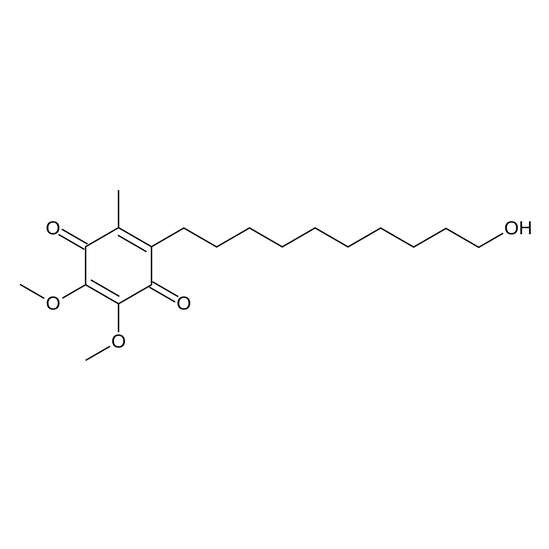
| CAS Number | 58186-27-9 |
| Other Names | Idebenona, Idebenonum |
| IUPAC Name |
2-(10-hydroxydecyl)-5,6-dimethoxy-3-methyl-cyclohexa-2,5-diene-1,4-dione
|
| Molecular Formula | C₁₉H₃₀O₅ |
| Molecular Weight | 338.44 |
| Purity | ≥99% Pure (LC-MS) |
| Liquid Availability | N/A |
| Powder Availability |  5 grams, 10 grams |
| Gel Availability | N/A |
| Storage | Store in cool dry environment, away from direct sunlight. |
| Terms | All products are for laboratory developmental research USE ONLY. Products are not for human consumption. |
What is Idenbenone?
Idebenone is a synthetic analog of coenzyme Q10 that acts as a nootropic compound due to its antioxidant properties and its role in supporting mitochondrial function. Originally developed to treat neurodegenerative conditions such as Alzheimer’s disease, idebenone has since been investigated for its cognitive enhancing effects, particularly in the context of age-related cognitive decline and neurological disorders. By improving cellular energy production and reducing oxidative stress in the brain, idebenone may help enhance memory, focus, and overall cognitive resilience. Current research regarding idebenone is focused on further defining the neuroprotective profile of the compound.
Main Research Findings
1) Idebenone administration was found to prevent ferroptosis through the inhibition of excessive autophagy of the ROS-AMPK-mTOR pathways in order to preserve cardiac function following myocardial infarction.
2) Treatment with idebenone was shown to reduce respiratory complications in patients with Duchenne’s muscular dystrophy.
Selected Data
1) This study conducted by the research team of Li et al investigates the effects of idebenone in both in vitro and in vivo models to explore its potential protective effects, particularly under conditions of oxidative stress and ferroptosis. The in vitro component utilized H9c2 cells derived from embryonic rat heart-derived cardiomyocytes, while the in vivo portion employed a mouse model of myocardial infarction (MI).
H9c2 cells were cultured in DMEM supplemented with 10% fetal bovine serum. For experimentation, the cells were seeded and cultured for 24 hours before being treated with various compounds. To model oxidative stress, cells were exposed to different concentrations of hydrogen peroxide, ranging from 1 to 400 μmol/L, for 4 hours. Based on preliminary results, 100 μmol/L hydrogen peroxide was selected for subsequent experiments. Idebenone at concentrations of 0.2, 0.5, 1, and 2 μmol/L was administered 2 hours prior to hydrogen peroxide treatment. Other agents such as deferoxamine in doses of 100 μmol/L, ferrostatin-1 in doses of 12 μmol/L, and metformin in doses of 10 mmol/L)were also used to assess their interaction with idebenone; for instance, metformin was added 0.5 hours before idebenone treatment [1].
To induce ferroptosis, cells were treated with various concentrations of RSL3 ranging from 0.1 to 10 μmol/L,for 3 hours. From these dosages, 0.5 μmol/L RSL3 was chosen for future testing. Idebenone was again administered 2 hours prior to RSL3 exposure. Rapamycin in a 100 nmol/L dose was also included in some experiments, and was added 0.5 hours before idebenone at 1 μmol/L.
To evaluate cell viability, H9c2 cells were seeded in 96-well plates and incubated for 24 hours. Following drug treatment, a Cell Counting Kit-8 assay was used. Medium was replaced with DMEM containing 10% CCK-8 reagent, and absorbance at 450 nm was measured after 1 hour of incubation. Control wells containing only medium were used to eliminate background interference, and untreated cells were considered 100% viable. Additionally, a LIVE/DEAD Viability/Cytotoxicity assay was used for visualization. In this test, cells were seeded in 24-well plates, treated with drugs, and incubated with 1.5 μM calcein AM and 3 μM ethidium homodimer-1 for 40 minutes. Fluorescence microscopy revealed green for live cells and red for dead cells [1].
Intracellular ROS levels were measured using a ROS Assay Kit. After seeding and drug treatment, H9c2 cells were incubated with diluted DCFH-DA for 20 minutes, followed by staining for 10 more minutes. Fluorescent microscopy was used to observe the cells. Additionally, ROS production was measured in heart species using dihydroethidium staining. Frozen tissue sections were incubated in ROS dye for 30 minutes and then with DAPI for nuclear staining and fluorescent microscopy was used for imaging [1].
Lipid peroxidation was assessed using Liperfluo, a fluorescent probe specific to lipid peroxides. Cells were incubated with 5 μM Liperfluo for 30 minutes, followed by 10 minutes with Hoechst 33342. After washing, fluorescence microscopy was used to detect lipid peroxidation. Furthermore, ATP content was measured in H9c2 cells using an ATP assay kit. Cells were cultured in 6-well plates and treated with idebenone and hydrogen peroxide. After treatment, cells were lysed, and the lysate was centrifuged. The supernatant was collected for ATP analysis. Protein concentrations were determined using the BCA assay, and ATP levels were calculated from a standard curve based on luminescence readings.
For the in vitro portion of the study, male mice were randomly divided into three groups including: sham-operated with vehicle, myocardial infarction with vehicle, and myocardial infarction with idebenone. Idebenone, at a dose of 100 mg/kg, or vehicle was administered via intraperitoneal injection 24 hours and 2 hours before surgery. Myocardial infarction was induced by ligation of the left anterior descending coronary artery, confirmed by pallor in the heart tissue. Sham-operated mice underwent the same procedure without left anterior descending ligation. Post-surgery, idebenone or vehicle was administered once daily for either 3 days or 4 weeks. After the treatment period, hearts were collected for analysis including iron content, levels of reactive oxygen species , malondialdehyde, transmission electron microscopy, and immunofluorescence [1].
Cardiac function was monitored by transthoracic echocardiography using the Vevo2100 imaging system. Measurements were taken at baseline, and 1 and 4 weeks post-surgery. A blinded cardiologist assessed cardiac parameters, including ejection fraction and fractional shortening, using software integrated into the ultrasound system. Heart tissues were fixed in 4% paraformaldehyde, embedded in paraffin, and sectioned into 5 μm slices for hematoxylin and eosin and Masson’s trichrome staining. These stains were used to observe general morphology and to quantify the area affected by myocardial infarction using light microscopy [1].
For ultrastructural analysis, heart tissue was quickly harvested and sectioned within minutes of excision. Samples were fixed in glutaraldehyde, post-fixed in osmium tetroxide, embedded in epoxy resin, and sectioned into 60–80 nm slices. After staining with uranyl acetate and lead citrate, sections were examined using a transmission electron microscope [1].
2) The study performed by researchers McDonald et al evaluated the efficacy and safety of idebenone in Duchenne muscular dystrophy (DMD) patients who were not receiving concomitant glucocorticoids. A total of 64 DMD patients were enrolled in the study, which compared daily treatment with 900 mg idebenone to a placebo treatment [2].
To be eligible for the study, patients had to have a baseline peak expiratory flow percent predicted of ≤80%, indicating compromised pulmonary function. The treatment duration was set at 52 weeks, and the intention-to-treat population consisted of 31 patients randomized to the idebenone group and 33 to the placebo group. Participants were between 10 and 18 years of age, with an average age of 14.3 years. Patients in the placebo group were slightly older on average with an average age of 15.0 years, compared to those in the idebenone group that had an average age of 13.5 years [2].
At the start of the trial, 92.2% patients were non-ambulatory, and this proportion was consistent between treatment groups affecting 90% of those in the idebenone group and 94% of those in the placebo group. 43.8% of the participants had never previously used glucocorticoids. Those who had used glucocorticoids in the past were required to have discontinued them at least 12 months prior to the start of the study. No patient was allowed to take glucocorticoids during the 52-week trial period.
Spirometry was used to assess respiratory function, with evaluations conducted at baseline and at weeks 13, 26, 39, and 52. These tests were administered by qualified and certified operators using the Pneumotrac Spirometer 6800. The primary measures of respiratory function were peak expiratory flow and forced vital capacity, which were adjusted for maturational growth by converting them into percent predicted values. Since many DMD patients experience growth challenges, body height used in these conversions was calculated using ulna length measurements [2].
A post-hoc efficacy analysis was conducted to evaluate treatment-emergent adverse events, with a focus on bronchopulmonary adverse events. These events were identified and evaluated by an independent, blinded pulmonary specialist. Bronchopulmonary adverse events were defined as treatment-emergent adverse events affecting the larynx, trachea, bronchi, lungs, or lower airways. In contrast, adverse events involving the nose, throat, or sinuses were excluded from the bronchopulmonary adverse events classification due to their less direct impact on lower respiratory health. The included bronchopulmonary adverse events were those labeled under preferred medical terms such as bronchitis, pneumonia, laryngitis, upper respiratory tract infection, influenza with respiratory symptoms, viral respiratory infections, cough, dyspnea, and respiratory failure, including acute respiratory failure. Additionally, treatment-emergent adverse events like nasopharyngitis, otitis media, rhinitis, allergic rhinitis, or sleep apnea syndrome were excluded from the bronchopulmonary adverse events category [2].
Only bronchopulmonary adverse events that occurred during the period of study medication intake were considered for the analysis. Event duration was calculated from the start to the end date as recorded in the safety database. If the bronchopulmonary adverse events extended beyond the medication period, only the duration during treatment was considered. Yearly incidence rates of bronchopulmonary adverse events were also calculated, normalized to patient-years of exposure to the study medication. This allowed for a fair comparison of event frequency relative to the amount of time patients were exposed to either idebenone or placebo.
Additionally, the use of systemic antibiotics was evaluated to gauge how frequently airway infections or related complications may have required treatment. Only antibiotics with systemic exposure were included in the analysis. The focus was on antibiotics used specifically for respiratory infections, with consideration of the indications reported in the study’s concomitant medication records. The antibiotics most commonly administered were oral formulations of amoxicillin or amoxicillin/clavulanic acid [2].
In conclusion, this portion of the DELOS trial report provides a detailed account of the patient population, methodology for assessing respiratory function, and procedures used to identify and analyze bronchopulmonary adverse events and antibiotic usage. These analyses were crucial in evaluating the efficacy and safety profile of idebenone in DMD patients, particularly its potential to mitigate respiratory decline and associated complications [2].
Discussion
1) The study conducted by the research team of Li et al investigated the protective effects of idebenone against hydrogen peroxide-induced oxidative damage, ferroptosis, and excessive autophagy in H9c2 cardiomyocytes and in a myocardial infarction (MI) mouse model. The aim was to determine whether idebenone, an antioxidant compound, can attenuate cell death, preserve mitochondrial function, and inhibit ferroptosis and autophagy under oxidative stress conditions [1].
To simulate oxidative stress, H9c2 cells were treated with hydrogen peroxide. A concentration of 100 μM was found to significantly reduce cell viability to 36.94% of the control group, and was thus used in subsequent experiments. Pretreatment with idebenone at concentrations of 0.5 μM and 1 μM notably improved cell viability by 59.80% and 53.07% of the control, respectively, with the 0.5 μM dose proving most effective. LIVE/DEAD assays confirmed that idebenone reduced the percentage of dead cells from 40.14% in the hydrogen peroxide group, to 18.74%, indicating a significant cytoprotective effect.
The researchers then explored whether hydrogen peroxide induced ferroptosis, a form of regulated cell death associated with iron accumulation and lipid peroxidation. Treatment with deferoxamine and ferrostatin-1, known ferroptosis inhibitors, alleviated damage induced by hydrogen peroxide, supporting the involvement of ferroptosis. Western blot analysis showed that hydrogen peroxide reduced the expression of GPX4, FPN1, and FTH1; all of which are key proteins involved in inhibiting ferroptosis, while idebenone pretreatment upregulated their expression by 1.44-fold, 1.51-fold, and 3.10-fold, respectively [1].
Further analysis demonstrated that hydrogen peroxide increased intracellular reactive oxygen species, lipid peroxidation, and lipofluor signal intensity. Idebenone pretreatment significantly reduced these effects, lowering reactive oxygen species production and lipid peroxidation levels by 0.52-fold and 0.37-fold, respectively. Idebenone also preserved mitochondrial function, as evidenced by increased ATP levels and improved mitochondrial membrane potential, measured using the JC-1 probe. Notably, ATP levels in the idebenone-only group exceeded those of the control, suggesting enhanced mitochondrial efficiency. Idebenone also reduced intracellular iron accumulation, confirmed by a 0.74-fold decrease in FerroOrange fluorescence [1].
Regarding autophagy, idebenone mitigated hydrogen peroxide-induced excessive autophagy in H9c2 cells. This was demonstrated by decreased LC3II/LC3I ratio and increased p62 expression by 0.42-fold and 2.19-fold compared to the hydrogen peroxide group, respectively. Transmission electron microscopy and ad-GFP-LC3B fluorescence assays revealed reduced numbers of autophagosomes in idebenone-treated cells compared to the hydrogen peroxide group, confirming that idebenone suppressed excessive autophagic activity.
To explore the broader regulatory networks involved, the study used computational biology techniques, including weighted gene co-expression network analysis and gene set variation analysis. Analysis of high-throughput data from atherosclerosis and MI patient samples revealed strong correlations among ferroptosis, autophagy, and AMPK-mTOR signaling pathways. Functional enrichment analysis indicated that these pathways were significantly activated in disease states. Specifically, MI patients exhibited elevated activity in ferroptosis, autophagy, and AMPK pathways, while mTOR signaling was inhibited. These findings suggest that dysregulation of these pathways contributes to ischemic events and may be targeted by therapeutic interventions like idebenone [1].
In vivo experiments further validated the cardioprotective effects of idebenone. Mice were divided into sham, MI + vehicle, and MI + idebenone groups. Idebenone was administered intraperitoneally before and after MI surgery. Echocardiographic analysis showed that MI significantly reduced left ventricular ejection fraction and fractional shortening. Idebenone treatment was found to improve ejection fraction by 38.55% at week 1 and 33.76% at week 4, while fractional shortening was improved by 18.35% at week 1 and 15.86% at week 4, compared to the MI + vehicle group. Histological analysis revealed that idebenone reduced infarct size from 21.35% in the MI group to 15.4% in the MI + idebenone group, indicating preserved myocardial integrity.
To assess the effects of idebenone on ferroptosis in vivo, the study measured reactive oxygen species, lipid peroxidation, and iron levels in myocardial tissues. Idebenone reduced reactive oxygen species levels to 0.72-fold and lipid peroxidation levels to 1.2-fold of the sham group, reversing the MI-induced elevations. Similarly, myocardial iron accumulation was decreased from 1.5-fold to 1.2-fold following idebenone treatment. Western blot results showed that idebenone restored the expression of GPX4, FTH1, and FPN1 proteins, which had been suppressed by MI [1].
Autophagy markers were also assessed in heart tissues. MI increased the LC3II/LC3I ratio by 3.5-fold and decreased p62 levels by 0.6-fold, indicating excessive autophagy. Idebenone attenuated these changes, reducing the LC3II/LC3I ratio to 1.5-fold and increasing p62 to 0.9-fold compared to the sham group. Transmission electron microscopy and LC3 immunofluorescence assays confirmed that idebenone reduced autophagosome numbers significantly in MI mice [1].
In summary, idebenone effectively protects against oxidative stress-induced damage in both in vitro and in vivo models. It alleviates hydrogen peroxide-induced cell death, inhibits ferroptosis by reducing reactive oxygen species, lipid peroxidation, and iron accumulation, as well as restores mitochondrial function. Additionally, idebenone prevents excessive autophagy and regulates the AMPK-mTOR pathway, which is crucial in MI pathophysiology. These findings highlight idebenone’s potential as a therapeutic agent for ischemic heart diseases by targeting oxidative stress, ferroptosis, and autophagy [1].
2) The experimental trial conducted by the research team of McDonald et al investigated the effect of idebenone on respiratory function and bronchopulmonary complications in patients with Duchenne muscular dystrophy. A key objective was to determine whether idebenone could help prevent patients from falling below clinically significant thresholds in peak cough flow and forced vital capacity, which are important indicators of respiratory health in DMD.
In a pre-specified analysis, the proportion of patients whose peak cough flow dropped below the threshold of 160 L/min was assessed. At baseline, 26 patients in the idebenone group and 33 in the placebo group had peak cough flow above this threshold. During the 52-week treatment period, only 3.8% of patients in the idebenone group dropped below the threshold, compared to 18.2% in the placebo group. Similarly, the study analyzed the number of patients whose forced vital capacity fell below 1 L, another clinically relevant benchmark. Of the patients who started the study with forced vital capacity between 1 and 1.5 L, 55.6% of patients in the placebo group experienced a decline below 1 L, whereas only 16.7% in the idebenone group did. Across the entire intent-to-treat population, 15.6% of placebo patients fell below this threshold versus only 3.2% in the idebenone group. These findings suggest idebenone helps delay or prevent declines in critical respiratory function levels [2].
The trial also assessed bronchopulmonary adverse events, which are often associated with compromised respiratory function and pose serious health risks in DMD. A total of 19.4% of patients in the idebenone group reported 7 bronchopulmonary adverse events, whereas 51.5% of patients in the placebo group reported 28 bronchopulmonary adverse events. Further analysis using a Cox Proportional Hazards model showed that the hazard ratio for experiencing at least one bronchopulmonary adverse event was 0.327, in favor of idebenone. Additionally, a Proportional Means Regression Model considering all bronchopulmonary adverse events indicated a hazard ratio of 0.281, again favoring idebenone [2].
Although the mean duration of bronchopulmonary adverse events was slightly longer in the idebenone group, the median duration was similar between groups. The total number of bronchopulmonary adverse events days was significantly lower for idebenone at 82 days, compared to placebo at 222 days, indicating fewer and shorter events in the idebenone group. Hospitalizations due to respiratory causes were more frequent in the placebo group, with 4 patients admitted for issues like acute respiratory failure and pneumonia, compared to only 1 patient in the idebenone group, who was hospitalized for sleep apnea syndrome. These data consistently support the conclusion that idebenone reduces both the frequency and severity of bronchopulmonary complications in DMD patients.
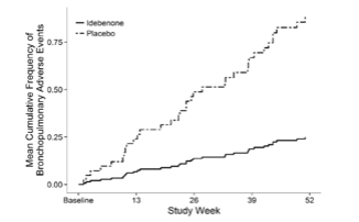
Figure 1: The average cumulative frequency of bronchopulmonary adverse events across 52 study weeks in the placebo-treated group and idebenone-treated group.
Further analyses were conducted to evaluate the influence of age, baseline respiratory function, and prior glucocorticoid use on the occurrence of bronchopulmonary adverse events. There were no major age differences between patients with or without bronchopulmonary adverse events within each treatment group. A slight age imbalance was noted, with the placebo group generally being older, but this did not account for the difference in outcomes. Baseline respiratory function, measured by peak expiratory flow, predicted peak expiratory flow, forced vital capacity, and predicted forced vital was largely similar between groups, particularly for patients who did not experience bronchopulmonary adverse events. However, among those who did experience bronchopulmonary adverse events, placebo patients had lower predicted forced vital capacity at baseline than idebenone-treated patients, suggesting that worse baseline function might increase susceptibility to complications in the absence of idebenone [2].
A multivariate Cox regression model incorporating age, predicted peak expiratory flow, predicted forced vital capacity, and prior glucocorticoid usage, showed that none of these factors significantly influenced bronchopulmonary adverse events risk, indicating that the reduction in bronchopulmonary adverse events was primarily due to idebenone treatment rather than baseline characteristics. The Proportional Means Regression model yielded similar findings.
Further stratification based on baseline pulmonary function revealed that the protective effects of idebenone were especially pronounced in patients with more severe impairment. Among patients with baseline predicted forced vital capacity or peak expiratory flow of ≤ 60%, those in the placebo group experienced over one bronchopulmonary adverse event per year on average, while idebenone-treated patients experienced substantially fewer or no bronchopulmonary adverse events. Notably, none of the idebenone-treated patients with baseline values ≤ 40% reported any bronchopulmonary adverse events, whereas placebo-treated patients with similar impairments did, again underscoring idebenone’s benefit in high-risk individuals [2].
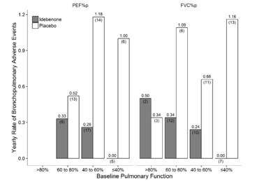
Figure 2: Yearly rate of bronchopulmonary adverse events measured by predicted peak expiratory volume and predicted forced vital capacity in the idebenone-treated and placebo-treated groups
The study also assessed systemic antibiotic use, which often accompanies severe respiratory infections. In the idebenone group, 22.6% of patients reported 8 antibiotic use episodes, compared to 39.4% of patients and 17 episodes in the placebo group. While the average duration of antibiotic use was similar across groups, total antibiotic days were fewer in the idebenone group at 65 days versus placebo at 105 days. Though neither the time to first antibiotic use nor the cumulative frequency reached statistical significance, hazard ratios from both the Cox model and the Proportional Means Regression model favored idebenone [2].
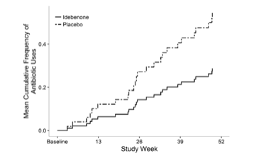
Figure 3: The average cumulative frequency of antibiotic groups across 52 study weeks in placebo-treated and idebenone-treated groups.
In summary, the trial provides significant evidence that idebenone treatment in DMD patients can slow the decline in respiratory function, reduce the incidence and severity of bronchopulmonary complications, and lessen the need for antibiotics. These benefits were most notable in patients with more advanced respiratory impairment, reinforcing the clinical value of idebenone in this high-risk population [2].
Disclaimer
**LAB USE ONLY**
*This information is for educational purposes only and does not constitute medical advice. THE PRODUCTS DESCRIBED HEREIN ARE FOR RESEARCH USE ONLY. All clinical research must be conducted with oversight from the appropriate Institutional Review Board (IRB). All preclinical research must be conducted with oversight from the appropriate Institutional Animal Care and Use Committee (IACUC) following the guidelines of the Animal Welfare Act (AWA).
Citations
[1] Li D, Zhang G, Wang Z, et al. Idebenone attenuates ferroptosis by inhibiting excessive autophagy via the ROS-AMPK-mTOR pathway to preserve cardiac function after myocardial infarction. Eur J Pharmacol. 2023;943:175569. doi:10.1016/j.ejphar.2023.175569
[2] McDonald CM, Meier T, Voit T, et al. Idebenone reduces respiratory complications in patients with Duchenne muscular dystrophy. Neuromuscul Disord. 2016;26(8):473-480. doi:10.1016/j.nmd.2016.05.008
Idebenone is sold for laboratory research use only. Terms of sale apply. Not for human consumption, nor medical, veterinary, or household uses. Please familiarize yourself with our Terms & Conditions prior to ordering.


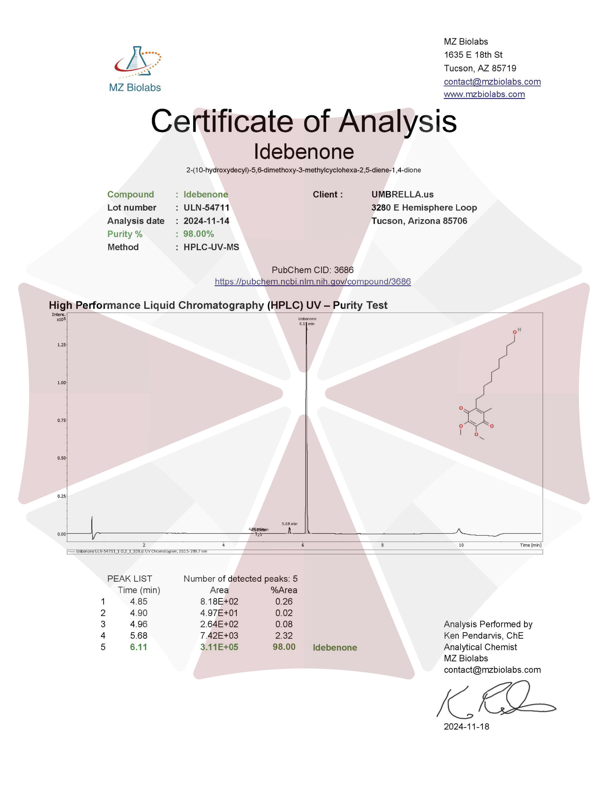
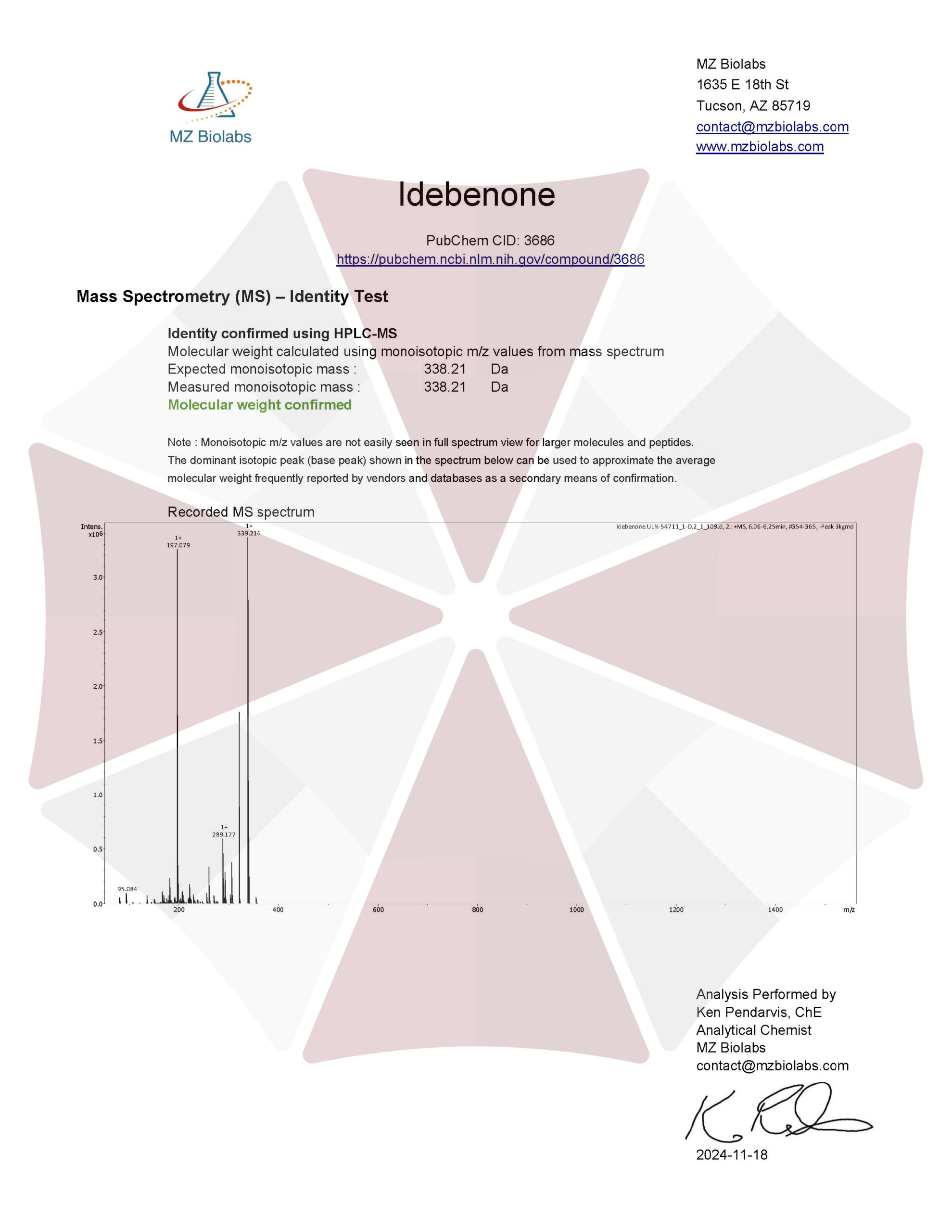
| File Name | View/Download |
| 2024-11-14-Umbrella-Labs-Idebenone-Certificate-Of-Analysis-COA.pdf |  |
VIEW CERTIFICATES OF ANALYSIS (COA)

Reviews
There are no reviews yet.