


Description
Mitoquinone Mesylate Nootropic Liquid








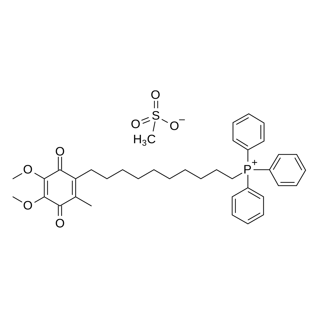
| CAS Number | 845959-50-4 |
| Other Names | Mitoquinone methanesulfonate, MitoQ, Mitoquinone (mesylate), UNII-6E01CG547T, 6E01CG547T, MitoQ10 mesylate, Mitoubiquinone mesylate, SCHEMBL4423673, CHEMBL4074884, DTXSID00233573 |
| IUPAC Name |
10-(4,5-dimethoxy-2-methyl-3,6-dioxocyclohexa-1,4-dien-1-yl)decyl-triphenylphosphanium;methanesulfonate
|
| Molecular Formula | C₃₈H₄₇O₇PS |
| Molecular Weight | 678.8 |
| Purity | ≥99% Pure (LC-MS) |
| Liquid Availability |  30mL liquid (20mg/mL, 600mg bottle) |
| Powder Availability | 1 gram |
| Storage | Store in cool dry environment, away from direct sunlight. |
| Terms | All products are for laboratory developmental research USE ONLY. Products are not for human consumption. |
What is Mitoquinone Mesylate?
Mitoquinone mesylate (MitoQ) is a novel mitochondria-targeted antioxidant that has emerged as a promising compound for supporting cognitive function and promoting healthy aging. MitoQ is composed of a coenzyme Q10 derivative attached to a lipophilic triphenylphosphonium ion, enabling it to penetrate cell membranes and accumulate selectively within mitochondria. This targeted delivery allows MitoQ to neutralize reactive oxygen species at their primary site of production, thereby reducing oxidative stress, preserving mitochondrial integrity, and enhancing cellular energy production. Because mitochondrial dysfunction and oxidative damage are closely associated with cognitive decline and neurodegenerative diseases, MitoQ has been extensively investigated for its neuroprotective potential. Preclinical studies have demonstrated improvements in neuronal survival, synaptic function, learning, and memory, while also showing reductions in neuroinflammation and mitochondrial damage. Available evidence suggests that MitoQ is well tolerated and may provide therapeutic benefits in disorders characterized by impaired mitochondrial function. These findings have positioned MitoQ as a promising nootropic candidate with potential applications in maintaining cognitive performance and reducing the impact of age-related neurological decline.
Main Research Findings
1) Treatment with mitoquinone mesylate was shown to reduce pathological features of allergic asthma in both lean and obese animal models.
2) Mitoquinone mesylate decreased brain inflammation in an animal model of chronic HIV infection.
Selected Data
1) The study by Chandrasekaran et al was designed to investigate whether mitoquinone mesylate (MitoQ), a mitochondria-targeted antioxidant, could reduce the pathological features of allergic asthma in both lean and obese mice. Because obesity is associated with more severe asthma, increased oxidative stress, mitochondrial dysfunction, and reduced responsiveness to conventional therapies, the investigators hypothesized that selectively reducing mitochondrial reactive oxygen species (mROS) would attenuate airway inflammation, remodeling, and airway hyperresponsiveness. To test this hypothesis, the researchers employed a comprehensive experimental design that combined in vitro cell culture experiments, diet-induced obesity mouse models, histological analyses, proteomics, molecular biology, and physiological assessments of lung function. The study also sought to determine whether the beneficial effects of MitoQ resulted specifically from its antioxidant ubiquinone component or from its lipophilic triphenylphosphonium (dTPP⁺) carrier moiety, which independently accumulates within mitochondria [1].
The in vitro component of the study utilized primary normal human bronchial epithelial (pHBE) cells cultured in specialized airway epithelial growth medium. After an initial adaptation period, epithelial cells were exposed to house dust mite (HDM) extract, one of the most common environmental allergens responsible for allergic asthma. HDM was administered at a concentration of 25 μg/mL to induce an inflammatory response, while control cells received phosphate-buffered saline. Twenty-four hours later, experimental groups were treated with either MitoQ or decyl-triphenylphosphonium (dTPP⁺), an inactive structural analog of MitoQ lacking the antioxidant ubiquinone component. Cells received a second dose of HDM together with the antioxidant treatments before culture supernatants and cellular lysates were collected for cytokine measurements and protein analyses. All cell culture experiments were performed in triplicate and independently repeated on multiple occasions to ensure reproducibility of the findings [1].
To model allergic asthma in vivo, the investigators used male C57BL/6NJ mice because this strain readily develops obesity when maintained on a high-fat diet. Four-week-old mice were randomly assigned to either a low-fat diet containing 10% of calories from fat or a high-fat diet containing 60% of calories from fat for twenty weeks. This dietary intervention produced lean and obese experimental groups that allowed investigators to compare the effects of obesity on allergic airway disease.
After completion of the dietary period, mice were divided into three treatment groups consisting of phosphate-buffered saline controls, HDM-challenged mice, and HDM-challenged mice receiving MitoQ. Allergic airway disease was induced through intranasal sensitization with 25 μg of HDM on experimental days one and seven, followed by repeated HDM challenges on days fourteen through eighteen. Mice assigned to the treatment group received intraperitoneal injections of MitoQ at a dose of 6 mg/kg on days seventeen through nineteen, while additional experiments administered equimolar concentrations of dTPP⁺ to determine whether the antioxidant or carrier component mediated therapeutic effects. Animals were euthanized on day twenty to evaluate acute inflammatory responses, while separate cohorts were examined on day twenty-seven to investigate the persistence and resolution of airway inflammation.
Following completion of the treatment protocol, bronchoalveolar lavage fluid (BALF) and lung tissue were collected for comprehensive assessment of airway inflammation and remodeling. Bronchoalveolar lavage was performed through tracheal cannulation using phosphate-buffered saline, allowing investigators to quantify total inflammatory cells and identify specific immune cell populations including macrophages, neutrophils, eosinophils, and lymphocytes using both cytospin preparations and flow cytometry. Flow cytometric analyses employed established cell surface markers to distinguish inflammatory cell populations, while lung tissue homogenates were prepared for enzyme-linked immunosorbent assays (ELISAs) measuring inflammatory mediators including interleukin-1β (IL-1β), interleukin-33 (IL-33), eotaxin (CCL11), CCL20, eosinophil peroxidase (EPX), transforming growth factor-beta (TGF-β), periostin, and additional cytokines associated with allergic airway inflammation. Lactate dehydrogenase activity and dead-cell protease assays were also performed on bronchoalveolar lavage samples to evaluate cellular injury and cytotoxicity [1].
Histological analyses were conducted using paraffin-embedded lung tissue sections to evaluate structural changes associated with allergic asthma. Masson’s trichrome staining was performed to quantify collagen deposition and airway fibrosis, while Periodic acid-Schiff (PAS) staining was used to assess goblet cell hyperplasia and mucus production. Eosinophil infiltration within lung tissue was evaluated using immunohistochemical staining for eosinophil peroxidase (EPX), and stained sections were analyzed quantitatively using digital imaging software. Importantly, all histological scoring was performed independently by three blinded investigators to minimize observer bias. In addition to tissue staining, quantitative reverse-transcription polymerase chain reaction (qRT-PCR) was used to measure expression of genes associated with allergic inflammation and mucus production, including Il4, Gob5, Agr2, and α-SMA, while Western blotting evaluated activation of proteins involved in endoplasmic reticulum stress and unfolded protein response signaling, including phospho-IRE1, phospho-PERK, phospho-eIF2α, ATF6, and CHOP [1].
To investigate oxidative stress mechanisms, the investigators employed several specialized biochemical and proteomic techniques. Hyperoxidation of mitochondrial peroxiredoxin-3 (PRX3), an established indicator of mitochondrial reactive oxygen species production, was measured using immunoprecipitation followed by Western blot analysis. To identify oxidized proteins throughout the lung, proteins containing cysteine sulfenylation modifications were selectively labeled using the dimedone-based probe DCP-Bio1 and isolated using streptavidin affinity purification. Purified proteins were digested with trypsin and analyzed by high-resolution liquid chromatography-tandem mass spectrometry (LC-MS/MS). Protein identification was performed using the SEQUEST search algorithm against the UniProt mouse protein database, while quantitative spectral counting allowed comparison of oxidized proteins between lean, obese, HDM-treated, and MitoQ-treated animals. These analyses enabled investigators to characterize the pulmonary “oxidome” and identify proteins whose oxidative modification was altered by allergic inflammation and antioxidant therapy.
Functional respiratory outcomes were evaluated by measuring airway hyperresponsiveness (AHR). Anesthetized and mechanically ventilated mice underwent methacholine challenge testing, during which increasing concentrations of aerosolized methacholine were administered to provoke airway constriction. Three complementary physiological parameters were measured, including central airway resistance (Rn), tissue damping (G), and tissue elastance (H), providing a comprehensive assessment of airway mechanics. By integrating cellular experiments, animal models, pulmonary physiology, histopathology, molecular biology, and advanced proteomics, the investigators established a rigorous experimental framework for evaluating the therapeutic potential and molecular mechanisms of MitoQ in both lean and obesity-associated allergic asthma [1].
2) The study by Satta et al was designed to investigate whether MitoQ, a mitochondria-targeted antioxidant, could reduce brain inflammation in a humanized mouse model of chronic HIV infection receiving suppressive antiretroviral therapy (ART). Although modern ART effectively suppresses viral replication and prolongs survival in people living with HIV, many patients continue to develop HIV-associated neurocognitive disorders (HAND), which remain among the most common neurological complications of chronic HIV infection. Persistent viral reservoirs and chronic immune activation are believed to contribute to ongoing oxidative stress, mitochondrial dysfunction, neuroinflammation, and neuronal injury despite successful viral suppression. Because mitochondria are a major source of reactive oxygen species (ROS) within the brain, the investigators hypothesized that selectively reducing mitochondrial oxidative stress with MitoQ would decrease inflammatory signaling and attenuate neuroinflammation in a preclinical model of chronic treated HIV infection. The primary objective of the study was therefore to determine whether long-term MitoQ administration could reduce proinflammatory cytokine and chemokine production within the brains of HIV-infected humanized mice maintained on effective ART [2].
To investigate this hypothesis, the researchers utilized NOD scid gamma (NSG) Bone Marrow-Liver-Thymic (BLT) humanized mice, a well-established experimental model that supports human immune cell development and HIV infection while closely reproducing important aspects of human HIV immunopathogenesis. Equal numbers of male and female mice were included to minimize potential sex-related differences in inflammatory responses. In total, twenty-seven humanized mice were generated according to previously validated protocols and maintained under institutional and federally approved animal care guidelines. Humanized BLT mice possess functional human immune cells capable of supporting HIV replication and therefore provide an appropriate platform for evaluating persistent immune activation during chronic infection despite antiretroviral therapy.
After successful engraftment, the mice were experimentally infected with the dual-tropic HIV-1 89.6 viral strain to establish chronic HIV infection. Once infection was established, all infected animals received combination antiretroviral therapy consisting of tenofovir disoproxil fumarate (8.75 mg/kg), emtricitabine (13 mg/kg), and raltegravir (17.5 mg/kg). These medications were administered daily throughout the experimental period to achieve sustained suppression of viral replication. The antiretroviral regimen had previously been validated by the investigators and closely resembles combination therapy used clinically to control HIV infection. The purpose of maintaining effective ART throughout the study was to reproduce the clinical situation in which patients exhibit well-controlled plasma viral loads but continue to experience chronic neuroinflammation and are therefore at risk for HIV-associated neurocognitive disorders [2].
To evaluate the therapeutic effects of mitochondrial antioxidant treatment, MitoQ was administered orally through the drinking water at a concentration of 500 μmol/L. Mice received continuous MitoQ supplementation for approximately ninety days while remaining on suppressive antiretroviral therapy. Control animals received flavored drinking water without MitoQ. Three experimental groups were established for comparison. Group A consisted of five uninfected humanized mice that served as healthy controls. Group B included twelve HIV-infected mice receiving antiretroviral therapy alone, representing chronic treated HIV infection without antioxidant intervention. Group C consisted of ten HIV-infected mice receiving both ART and MitoQ supplementation, allowing investigators to directly evaluate whether mitochondrial antioxidant therapy reduced neuroinflammation beyond the effects achieved with antiretroviral treatment alone. This three-group design enabled comparisons between healthy animals, chronically infected treated animals, and infected animals receiving adjunctive antioxidant therapy.
At the conclusion of the treatment period, brain tissue was collected from every animal for detailed immunological analysis. The primary outcome measure consisted of quantifying inflammatory cytokines and chemokines within brain tissue as biomarkers of neuroinflammation. Both human-derived cytokines produced by engrafted human immune cells and murine cytokines produced by resident mouse cells were evaluated to characterize inflammatory responses originating from multiple cellular sources. Cytokine concentrations were measured using highly sensitive multiplex Luminex immunoassays according to the manufacturer’s standardized protocols. This technology enabled simultaneous quantification of numerous inflammatory mediators from relatively small tissue samples while providing highly sensitive and reproducible measurements. Protein concentrations were normalized to total tissue protein determined by bicinchoninic acid (BCA) protein assay to account for differences in tissue sample size and protein content between animals [2].
The investigators measured several proinflammatory cytokines and chemokines previously implicated in the pathogenesis of HIV-associated neurocognitive disorders. Human inflammatory markers included interleukin-1β (IL-1β), interleukin-6 (IL-6), interleukin-8 (IL-8), tumor necrosis factor-alpha (TNF-α), CCL2, CCL3, CCL5, CXCL10, and the anti-inflammatory cytokine IL-10. Equivalent murine cytokines, including IL-1β, IL-6, TNF-α, IL-10, CCL2, CCL3, CCL5, and CXCL10, were also quantified to evaluate inflammatory signaling originating from resident mouse brain cells such as microglia and other innate immune cells. By simultaneously measuring both human and murine inflammatory mediators, the investigators were able to distinguish immune responses contributed by the engrafted human immune system from those generated by the murine central nervous system environment [2].
Throughout the study, suppression of HIV replication was monitored to ensure that observed changes in neuroinflammation were attributable to MitoQ treatment rather than differences in viral control. As reported previously by the investigators, plasma viremia was effectively suppressed in all HIV-infected animals after four weeks of combination ART, thereby creating a stable model of chronic treated HIV infection that closely resembles virologically suppressed patients receiving long-term antiretroviral therapy. Because viral replication remained controlled across infected groups, subsequent comparisons focused specifically on differences in inflammatory biomarker expression within brain tissue.
Cytokine concentrations were expressed as protein levels normalized to tissue protein content, and data were presented as box-and-whisker plots displaying minimum, median, and maximum values, with each individual data point representing one mouse. This statistical approach allowed investigators to determine whether adjunctive MitoQ therapy significantly reduced brain inflammation in HIV-infected humanized mice receiving effective antiretroviral treatment. Overall, the study combined a clinically relevant humanized mouse model, long-term combination antiretroviral therapy, targeted mitochondrial antioxidant supplementation, and comprehensive multiplex cytokine analysis to evaluate the potential of MitoQ as a novel therapeutic strategy for reducing chronic neuroinflammation associated with HIV infection [2].
Discussion
1) The study by Chandrasekaran et al demonstrated that MitoQ significantly attenuated multiple pathological features of allergic asthma in both lean and obese mice, supporting the hypothesis that targeting mitochondrial oxidative stress can improve airway inflammation and remodeling. Although both lean and obese mice developed allergic airway disease following repeated HDM exposure, obese animals exhibited a more severe disease phenotype characterized by increased tissue eosinophilia, elevated eotaxin production, greater airway hyperresponsiveness, and enhanced oxidative protein damage. MitoQ treatment substantially improved these pathological changes without affecting overall body weight or visceral adiposity, indicating that its therapeutic effects were independent of alterations in obesity itself. Throughout the study, MitoQ consistently reduced inflammatory responses, structural airway remodeling, oxidative stress, and physiological airway dysfunction in both dietary groups, although several benefits were particularly pronounced in obese allergic mice [1].
Analysis of bronchoalveolar lavage fluid demonstrated that HDM exposure produced robust airway inflammation in both lean and obese animals. Total inflammatory cell counts, neutrophils, eosinophils, and lymphocytes increased significantly after allergen challenge, while macrophage numbers remained relatively unchanged. MitoQ treatment selectively reduced lymphocyte accumulation in bronchoalveolar lavage fluid without substantially altering total inflammatory cells, neutrophils, or eosinophils.
Examination of inflammatory mediators within lung tissue revealed distinct differences between lean and obese animals. Lean mice exhibited greater increases in IL-1β and IL-33, whereas obese mice produced significantly higher concentrations of CCL11, a potent eosinophil chemoattractant. MitoQ significantly reduced IL-1β, IL-33, and CCL20 concentrations in both lean and obese mice while specifically decreasing eotaxin levels in obese animals. Expression of the Th2 cytokine Il4 also declined following treatment, particularly in lean mice. These findings demonstrated that MitoQ broadly suppressed allergic inflammatory signaling while exerting particularly beneficial effects on eosinophil-associated pathways that characterize obesity-related asthma [1].
One of the most important observations was the marked reduction in tissue eosinophilia produced by MitoQ. Although bronchoalveolar lavage eosinophil counts were similar between lean and obese HDM-treated mice, immunohistochemical staining revealed substantially greater eosinophil accumulation within the peribronchial and perivascular lung tissue of obese mice. This increase was confirmed by elevated EPX staining and higher tissue EPX concentrations. MitoQ significantly reduced both tissue eosinophil infiltration and EPX levels in obese allergic mice while having little effect on interleukin-5 concentrations, suggesting that its actions primarily involved reducing eosinophil recruitment rather than altering eosinophil differentiation.
Additional experiments examining persistent inflammation seven days after allergen exposure demonstrated that obese mice experienced delayed resolution of eosinophilic inflammation compared with lean animals. MitoQ accelerated resolution of both eosinophilic and lymphocytic inflammation during this later phase, indicating that the compound not only suppressed acute inflammatory responses but also promoted recovery following allergen exposure. These findings closely resemble clinical observations that obese individuals with asthma often exhibit increased tissue eosinophilia despite relatively modest eosinophil counts within the airway lumen [1].
MitoQ also substantially improved structural airway remodeling associated with chronic allergic inflammation. Histological analysis using Periodic acid-Schiff staining demonstrated that HDM exposure induced marked goblet cell metaplasia and mucus hypersecretion in both dietary groups, with particularly severe mucus accumulation observed in obese mice. MitoQ significantly reduced goblet cell hyperplasia in obese animals and suppressed expression of the mucus-associated genes Gob5 and Agr2, both of which are involved in mucin production and secretion. Similarly, Masson’s trichrome staining revealed extensive collagen deposition and inflammatory cell infiltration surrounding the airways after allergen challenge. MitoQ markedly reduced collagen accumulation, airway fibrosis, and peribronchial inflammatory infiltrates in both lean and obese mice. Consistent with these histological findings, concentrations of the profibrotic mediators TGF-β and periostin were significantly decreased following treatment, as was expression of α-SMA, a marker of airway smooth muscle remodeling. Collectively, these results demonstrated that MitoQ not only reduced inflammation but also limited the structural remodeling processes responsible for persistent airway dysfunction in allergic asthma [1].
Physiological measurements of lung function further confirmed the therapeutic benefits of MitoQ. Obese control mice exhibited greater baseline airway hyperresponsiveness than lean animals even before allergen exposure, reflecting the adverse effects of obesity on pulmonary physiology. HDM challenge further increased airway responsiveness in both groups, particularly central airway resistance in obese mice. Methacholine challenge testing demonstrated significant elevations in central airway resistance, tissue damping, and tissue elastance following allergen exposure. MitoQ significantly reduced airway resistance and tissue damping in both lean and obese mice, indicating improved airway function and reduced bronchoconstriction. Although changes in tissue elastance were less consistent, overall pulmonary function improved substantially following treatment. These findings represent important evidence that reducing mitochondrial oxidative stress can improve not only inflammatory biomarkers but also clinically relevant physiological measures of airway obstruction.
To investigate the mechanisms underlying these therapeutic effects, the investigators compared MitoQ with dTPP⁺, the lipophilic mitochondrial targeting moiety of MitoQ that lacks antioxidant activity. Surprisingly, dTPP⁺ reproduced many of the anti-inflammatory effects observed with MitoQ, including reductions in inflammatory cell infiltration, cytokine production, airway hyperresponsiveness, and markers of cellular injury. However, biochemical analysis demonstrated that dTPP⁺ actually increased mitochondrial reactive oxygen species production, whereas MitoQ reduced oxidation of mitochondrial PRX3, a sensitive marker of mitochondrial oxidative stress. These findings suggested that many anti-inflammatory effects of MitoQ were not mediated solely through direct scavenging of mitochondrial reactive oxygen species but instead involved additional mechanisms related to mitochondrial targeting or cellular signaling pathways influenced by the dTPP⁺ backbone [1].
Proteomic analyses provided additional mechanistic insight into MitoQ’s biological activity. Using selective labeling of cysteine-sulfenylated proteins followed by liquid chromatography-tandem mass spectrometry, investigators identified more than 1,100 proteins within the pulmonary oxidome. Obese HDM-treated mice demonstrated substantially greater protein oxidation than lean animals, particularly involving proteins associated with the ER and UPR. MitoQ significantly reduced oxidation of several ER-resident proteins, including GRP94, PDIA3, and GRP78, whereas dTPP⁺ failed to suppress these oxidative modifications. Western blot analyses further demonstrated activation of all three major UPR signaling pathways following allergen exposure. MitoQ significantly attenuated activation of each pathway in both lean and obese mice, while dTPP⁺ only partially inhibited IRE1 and PERK signaling. These findings indicate that MitoQ uniquely protects against oxidative modification of ER proteins and suppresses ER stress, providing a plausible mechanism linking mitochondrial oxidative stress to allergic airway inflammation and remodeling.
Overall, the investigators concluded that MitoQ effectively reduced allergen-induced airway inflammation, tissue eosinophilia, mucus hypersecretion, fibrosis, airway hyperresponsiveness, oxidative protein damage, and unfolded protein response activation in both lean and obese mouse models of allergic asthma. Although some anti-inflammatory effects appeared to arise from the dTPP⁺ carrier moiety, only MitoQ consistently reduced mitochondrial oxidative stress and prevented oxidation of critical endoplasmic reticulum proteins involved in unfolded protein response signaling. These findings suggest that mitochondrial oxidative stress and ER stress are important contributors to obesity-associated asthma and identify MitoQ as a promising therapeutic candidate for treating severe allergic asthma, particularly in obese individuals whose disease is characterized by excessive oxidative stress and persistent eosinophilic airway inflammation [1].
2) The study by the research team of Satta et al demonstrated that MitoQ significantly reduced neuroinflammation in a humanized mouse model of chronic HIV infection despite effective suppression of viral replication with combination ART. Although ART successfully controlled systemic HIV infection, persistent inflammatory activity remained evident within the brains of HIV-infected mice, reflecting the chronic immune activation that contributes to HAND. Administration of MitoQ markedly attenuated several key proinflammatory cytokines and chemokines implicated in neurodegeneration, providing the first preclinical evidence that a mitochondria-targeted antioxidant may reduce chronic brain inflammation associated with treated HIV infection. These findings support the hypothesis that mitochondrial oxidative stress continues to promote inflammatory signaling even after viral replication has been effectively suppressed and suggest that antioxidant therapy may represent a promising adjunctive treatment strategy for preventing or slowing the development of HAND [2].
Before evaluating the effects of MitoQ, the investigators first confirmed that the antiretroviral regimen effectively suppressed HIV replication throughout the study. Consistent with their previous investigations using the same humanized mouse model, all HIV-infected animals receiving combination ART achieved suppression of plasma viremia within four weeks of treatment. This finding was critical because it established that subsequent differences in inflammatory markers could not be attributed to uncontrolled viral replication but instead reflected persistent immune activation despite successful ART. The model therefore closely resembled the clinical situation observed in many individuals living with HIV, in whom viral replication remains well controlled while chronic inflammation and neurocognitive impairment continue to develop over time. By maintaining equivalent viral suppression across infected groups, the investigators created an experimental framework that isolated the specific effects of MitoQ on neuroinflammation independent of antiviral activity.
Analysis of brain tissue demonstrated that HIV infection continued to promote significant inflammatory responses despite long-term ART. Compared with uninfected control animals, HIV-infected mice receiving ART alone exhibited significantly elevated concentrations of several important human-derived inflammatory cytokines within the brain. Levels of human IL-1β, IL-8, and TNF-α were all significantly increased in the HIV-infected ART-treated group. Additionally, investigators observed a strong trend toward elevated concentrations of the chemokine human CCL2, although this increase narrowly missed conventional statistical significance. These findings demonstrated that substantial neuroinflammation persisted despite effective antiviral treatment, supporting previous evidence that suppression of circulating HIV does not completely eliminate inflammatory processes occurring within the central nervous system. Persistent activation of inflammatory cytokines such as IL-1β and TNF-α has been implicated in neuronal injury, synaptic dysfunction, and the cognitive impairment observed in patients with HIV-associated neurocognitive disorders [2].
The addition of MitoQ to suppressive antiretroviral therapy produced substantial reductions in multiple inflammatory mediators within the brain. Compared with HIV-infected mice receiving ART alone, animals treated with both ART and MitoQ demonstrated significantly lower concentrations of human IL-1β, IL-6, TNF-α, and CCL2. Each of these inflammatory markers declined significantly following MitoQ supplementation, indicating that mitochondrial antioxidant therapy effectively attenuated inflammatory signaling within the central nervous system. Among these biomarkers, IL-1β and TNF-α are recognized as major mediators of neurotoxicity and inflammasome activation, while IL-6 contributes to chronic neuroinflammation and CCL2 promotes recruitment of inflammatory immune cells into the brain. Simultaneous reductions in these cytokines therefore suggested that MitoQ broadly suppressed multiple inflammatory pathways rather than targeting only a single signaling mechanism. These findings strongly support the proposed role of mitochondrial reactive oxygen species in sustaining chronic neuroinflammation during treated HIV infection [2].
The investigators also evaluated inflammatory responses generated by resident murine cells within the brain to determine whether MitoQ influenced neuroinflammation originating from the host central nervous system. Similar to the changes observed for human cytokines, MitoQ significantly reduced murine IL-1β, IL-6, and TNF-α concentrations compared with HIV-infected animals receiving ART alone. These parallel reductions demonstrated that MitoQ attenuated inflammatory signaling arising from both engrafted human immune cells and endogenous murine brain cells, including microglia and other innate immune populations. The ability to simultaneously suppress inflammatory mediators from both cellular compartments suggests that mitochondrial oxidative stress contributes broadly to chronic neuroinflammatory processes regardless of cellular origin. Because activation of microglia represents a major pathological feature of HIV-associated neurocognitive disorders, these findings provide additional evidence that MitoQ may interrupt important mechanisms underlying neuronal injury during chronic treated HIV infection.
Not all inflammatory mediators were altered by treatment, however. The investigators found no significant differences among experimental groups in concentrations of either human or murine IL-10, CCL3, CCL5, or CXCL10. These results indicate that MitoQ selectively modulated specific proinflammatory signaling pathways without globally suppressing all cytokine production. Preservation of IL-10, an important anti-inflammatory cytokine, suggests that MitoQ did not broadly impair normal immune regulation. Likewise, the absence of significant changes in several chemokines indicates that the anti-inflammatory effects of MitoQ were relatively targeted toward cytokines closely associated with mitochondrial oxidative stress and inflammasome activation. This selective pattern of cytokine modulation may represent an important therapeutic advantage by reducing pathological inflammation while minimizing disruption of protective immune responses [2].
Based on these findings, the investigators concluded that MitoQ exerts both antioxidant and anti-inflammatory effects that may be particularly relevant for chronic HIV infection. They proposed that mitochondrial reactive oxygen species promote activation of the inflammasome, leading to increased production of IL-1β and other inflammatory mediators that contribute to neurotoxicity and cognitive decline. By selectively targeting mitochondria, MitoQ likely interrupts this cycle of oxidative stress and inflammatory activation, thereby reducing downstream cytokine production. The observed reductions in IL-1β, IL-6, TNF-α, and CCL2 are consistent with previous studies demonstrating anti-inflammatory effects of MitoQ in experimental models of multiple sclerosis, colitis, and other neurodegenerative disorders, further supporting a common mechanism involving suppression of mitochondrial oxidative stress and inflammasome activation.
Overall, this proof-of-concept study provided the first preclinical evidence that MitoQ can significantly reduce brain inflammation in a humanized mouse model of chronic HIV infection receiving suppressive antiretroviral therapy. Although the study focused exclusively on inflammatory biomarkers and did not directly assess cognitive function, neuronal survival, or behavioral outcomes, the substantial reductions in multiple cytokines and chemokines strongly support further investigation of MitoQ as an adjunctive therapy for HIV-associated neurocognitive disorders. The authors emphasized that MitoQ has been used safely as an oral dietary supplement and has demonstrated a favorable safety profile in previous human clinical trials, making it an attractive candidate for translation into clinical studies. They concluded that additional research should evaluate the effects of MitoQ on other mechanisms contributing to neuroinflammation and determine whether the reductions in inflammatory signaling observed in this study ultimately translate into improved neurological function and cognitive outcomes in people living with HIV [2].
Disclaimer
**LAB USE ONLY**
*This information is for educational purposes only and does not constitute medical advice. THE PRODUCTS DESCRIBED HEREIN ARE FOR RESEARCH USE ONLY. All clinical research must be conducted with oversight from the appropriate Institutional Review Board (IRB). All preclinical research must be conducted with oversight from the appropriate Institutional Animal Care and Use Committee (IACUC) following the guidelines of the Animal Welfare Act (AWA).
Citation
[1] Chandrasekaran R, Bruno SR, Mark ZF, et al. Mitoquinone mesylate attenuates pathological features of lean and obese allergic asthma in mice. Am J Physiol Lung Cell Mol Physiol. 2023;324(2):L141-L153. doi:10.1152/ajplung.00249.2022
[2] Satta S, Hugo C, Sharma M, et al. Mitoquinone mesylate attenuates brain inflammation in humanized mouse model of chronic HIV infection. AIDS. 2022;36(11):1609-1611. doi:10.1097/QAD.0000000000003291
Mitoquinone Mesylate is sold for laboratory research use only. Terms of sale apply. Not for human consumption, nor medical, veterinary, or household uses. Please familiarize yourself with our Terms & Conditions prior to ordering.


| File Name | View/Download |
| Coming Soon! |  |
VIEW CERTIFICATES OF ANALYSIS (COA)

Reviews
There are no reviews yet.