


Description
Cycloastragenol Nootropic Powder







| CAS Number | 78574-94-4 |
| Other Names | Cyclogalegigenin, Astramembrangenin |
| IUPAC Name |
24α,20-Epoxy-9,19-cyclo-9β-lanost-24-ene-3β,6α,16β,25-tetrol
|
| Molecular Formula | C₃₀H₅₀O₅ |
| Molecular Weight | 490.72 |
| Purity | ≥99% Pure (LC-MS) |
| Liquid Availability | N/A |
| Powder Availability |  N/A |
| Gel Availability | N/A |
| Storage | Store in cool dry environment, away from direct sunlight. |
| Terms | All products are for laboratory developmental research USE ONLY. Products are not for human consumption. |
What is Cycloastrogenol?
Cycloastragenol (CAG) is a triterpenoid saponin compound primarily isolated from the roots of Astragalus membranaceus. Gaining significant attention in recent years, this naturally occurring molecule is most notably recognized for its ability to activate telomerase, an enzyme responsible for maintaining and repairing telomeres, the protective caps at the ends of chromosomes. This mechanism has positioned cycloastragenol as a subject of considerable interest within the fields of anti-aging research, cellular senescence, and overall longevity.
Main Research Findings
1) When tested in lung cancer cell lines, cycloastragenol was shown to induce apoptosis and protective autophagy through activation of the AMPK/mTOR/ULK1 pathways.
2) Cycloastrogenol was shown to inhibit ROS-induced inflammasome activity in microglia resulting in reduced neuroinflammation and the promotion of microglia autophagy.
Selected Data
1) This study completed by the research team of Zhu et al investigated the anti-cancer effects of cycloastragenol (CAG) on human non-small cell lung cancer (NSCLC) cell lines and in an in vivo xenograft model, meticulously exploring the underlying molecular mechanisms, particularly apoptosis and autophagy pathways. The experimental design combined a wide array of in vitro cellular assays with in vivo animal studies, supported by detailed molecular analyses [1].
For in vitro experiments, two human NSCLC cell lines, H1299 and A549, were utilized. These were cultured in Dulbecco’s Modified Eagle’s Medium, supplemented with 10% fetal bovine serum and 1% penicillin-streptomycin solution. Cells were maintained in a controlled incubator at 37 °C with 5% carbon dioxide. Cycloastragenol, with a purity greater than 98% achieved by high-performance liquid chromatography, was dissolved in dimethyl sulfoxide (DMSO) for experimental use. Additional pharmacological modulators, including 3-methyladenine (3-MA), bafilomycin A1 (Baf-A1), chloroquine (CQ), and dorsomorphin (Com C), were obtained from Sigma-Aldrich to dissect specific cellular pathways.
Cell viability was assessed using an adenosine triphosphate (ATP) monitoring system, specifically the ATPLite luminescence assay kit. Cells were seeded in 96-well plates at a density of 1 × 10^3 cells/mL, treated with varying concentrations of CAG or DMSO, and then subjected to the luminescence assay to quantify ATP as an indicator of viable cells. Clonogenic survival assays further evaluated the long-term proliferative capacity by seeding 500 cells in 6-well plates, treating them, and then allowing them to grow for 14 days. Colonies were subsequently fixed and stained with 0.1% crystal violet before observation under an inverted microscope. Apoptosis induction was quantified using dual staining flow cytometry. H1299 and A549 cells were seeded in 6 cm dishes, treated with CAG or DMSO for 72 hours, and then stained with an Annexin V-fluorescein isothiocyanate (FITC)/propidium iodide (PI) apoptosis detection kit. Flow cytometry was then used to differentiate viable, early apoptotic, late apoptotic, and necrotic cells [1].
Molecular analyses relied heavily on Western blotting. Cell or tissue lysates were prepared using radioimmunoprecipitation assay buffer. Equal amounts of protein, quantified via bicinchoninic acid protein assay, were separated by sodium dodecyl-sulfate polyacrylamide gel electrophoresis (SDS-PAGE) and transferred to polyvinylidene difluoride (PVDF) membranes. Membranes were blocked with 5% nonfat milk and incubated with a comprehensive panel of primary antibodies targeting key proteins involved in apoptosis, autophagy, and associated signaling pathways. These included antibodies against total and phosphorylated forms of AMPK. Apoptosis markers included cleaved PARP and cleaved caspase 3. Autophagy markers comprised LC3B and Atg5. Other important proteins like NOXA, Bax, BIM, BID, and PUMA were also assessed. GAPDH and β-actin served as loading controls to ensure equal protein loading.
Immunofluorescence staining was used to visualize autophagy in A549 cells. Cells were fixed, permeabilized, and blocked before incubation with LC3B antibody. Nuclei were counterstained with DAPI, and images were captured using a fluorescence microscope. Transmission electron microscopy (TEM) provided ultrastructural details of autophagy. H1299 and A549 cells were fixed with glutaraldehyde and formaldehyde, post-fixed with osmium tetroxide, dehydrated, embedded in epoxy resin, and sectioned ultra-thinly. Sections were stained with heavy metals and examined using a TEM [1].
To investigate specific gene roles, small interfering RNA (siRNA) technology was employed for genetic knockdown. siRNA oligonucleotides targeting AMPK (siAMPK-1/2), ULK1 (siULK1-1/2), Atg5 (siAtg5-1/2), and NOXA (siNOXA-1/2) were produced. A non-specific control siRNA (siNC) and siβ-actin were also used. These siRNAs were transfected into A549 and H1299 cells using Lipofectamine 2000. Western blot was performed to validate the knockdown efficiency [1].
For in vivo validation, a subcutaneous tumor xenograft model was established using 4- to 6-week-old male Bagg Albino (BALB/c) nude mice. A549 cells were subcutaneously injected into the right axillae of mice. Approximately 5 days after tumor formation, mice were randomly assigned to either control or CAG treatment groups. The CAG group received daily intraperitoneal injections of 150 mg/kg of cycloastragenol, while the control group received vehicle. Mouse body weights were monitored every 4 days. After 17 days, all mice were euthanized, and tumors were collected, photographed, and weighed. Tumor growth was monitored by measuring length and diameter, from which tumor volume was calculated. Tissue sections from liver and kidney were collected for hematoxylin and eosin (H&E) staining to assess potential toxicity. Immunohistochemistry was performed on tumor tissues to detect Ki67, a proliferation marker, and LC3. Tumor protein lysates were also subjected to Western blotting to analyze the effects of CAG on key molecular targets in vivo [1].
2) This study completed by Feng et al investigated the neuroprotective effects of cycloastragenol (CAG) in Parkinson’s disease (PD) models, employing a comprehensive array of in vitro and in vivo methodologies. The research utilized a range of specialized chemicals and reagents to facilitate precise experimental control and accurate measurements. Crucial inhibitors and indicators included 3-MA and MitoSOX from MCE, and MitoQ. For the PD model, recombinant human α-synuclein with an A53T mutant (S-1002–2) and the adeno-associated virus vector AAV2/9-hSyn-SNCA(A53T)-α-Syn-WPRE were used. Cellular staining reagents included YO-PRO-1 (C2022) and DCFH-DA (S0033S). Furthermore, ELISA kits for detecting IL-18 and IL-1β were sourced utilized [2].
The in vivo component of the study established a PD mouse model using male C57BL/6 mice aged 8 weeks. Mice were randomly allocated into three groups: normal, model, and model+CAG. The normal group received an injection of AAV2/9-hSyn-vector-WPRE into the striatum, serving as a vehicle control. The model and model+CAG groups received stereotactic injections of AAV2/9-hSyn-SNCA(A53T)-α-Syn-WPRE into the striatum to induce α-synucleinopathy, a hallmark of PD. The injection parameters were standardized: 1 μl/side volume (4.5 × 10^13 gp/ml) delivered at a rate of 0.2 μl/min, using precise coordinates relative to bregma as follows: Ml ±2.0 mm, AP +0.8 mm, DV –2.2 mm. Four weeks post-AAV-α-Syn injection, mice in the model+CAG group began daily gavage administration of CAG at two different doses of 75 mg/kg or 125 mg/kg, for a duration of three months. The control groups (normal and model) received olive oil via gavage. Throughout the study, all mice were housed under controlled environmental conditions with temperature at 25 ± 1 ◦C, humidity at about 55 %, and 12-hour light/dark cycle, with free access to food and water [2].
Behavioral analyses were conducted during the light-on cycle from 10:00 to 16:00 to assess both motor and non-motor functions. Motor function was evaluated using standard tests such as the rotarod test, balance beam test, and gait test. Non-motor functions were assessed via the Open field test and the Elevated plus-maze. For advanced molecular profiling, quantitative 4D proteomics analysis was performed on brain samples. Brain tissues were first ground into cell powder, then lysed in a buffer containing 8 M urea and 1% protease inhibitor cocktail. For post-translational modification (PTM) analysis, the lysis buffer was supplemented with additional inhibitors, including 1% phosphatase inhibitor for phosphorylation and 50 mM NAM and 3 μM TSA for acetylation.
After centrifugation to remove debris, protein concentration was determined using a BCA kit. Proteins were subsequently digested with trypsin, and the resulting peptides were desalted using a C18 SPE column. The tryptic peptides were then dissolved in 0.5 M TEAB and labeled with specific TMT reagents before undergoing a 2-hour incubation. The labeled peptides were analyzed using a Liquid Chromatograph-Mass Spectrometer (LC-MS)/MS system, utilizing a nanoElute UHPLC system with a customized reversed-phase analytical column (25 cm long, 75/100 μm i.d.) and a timsTOF Pro mass spectrometer. Functional enrichment, clustering, and protein-protein interaction networks were analyzed through various annotations.
In vitro cellular studies involved primary microglial (PM) and primary neuron (PN) cultures. PM were cultured and harvested as previously reported, then planted onto poly-L-lysine-coated plates. PM received CAG treatment of 5 or 10 μM alone or in combination with 3-MA at a concentration of 5 mM, followed by α-Syn stimulation at 500 nM for 24 hr. PN cultures were prepared from cortical tissues of C57BL/6 mice. After membrane removal and digestion, neurons were transferred onto poly-L-lysine-precoated 24-well plates at a density of 2.5 × 10^4 cells/ml. Cultures were maintained in Neurobasal medium supplemented with penicillin/streptomycin and B27, with medium refreshment every 3 days. On the sixth day, PN were exposed to a 1:2 mixture of microglia-conditioned media (MCM) and neurobasal culture medium for 12 hours [2].
Additional cellular analyses included various staining techniques. Primary microglia were treated as described, and then stained with YO-PRO-1, Eth-D1, Hoechst, ROS (DCFH-DA), with images visualized under a confocal laser scanning microscope. ELISA was performed on primary microglial supernatant planted in 12-well plates to quantify IL-18 and IL-1β levels using commercial assay kits. Transmission electron microscopy (TEM) was used for ultrastructural analysis of cells, fixed in a solution of 2.5% glutaraldehyde and 2% paraformaldehyde, and processed according to established protocols. Immunocytochemistry (ICC) was applied to both brain slices and cells, processed as previously reported, and incubated with specific primary and corresponding secondary antibodies.
Gene expression analysis was conducted using real-time quantitative polymerase chain reaction (RT-qPCR). RNA was extracted from primary microglia, followed by cDNA synthesis. Amplification was performed with a real-time PCR system. The qPCR program involved denaturation, annealing/extension cycles, and a final step, with all experiments performed in triplicate. Relative mRNA expression was normalized against the housekeeping gene GAPDH. Western blotting was also utilized for quantitative immunoblotting of striatum tissues and cells, using primary antibodies [2].
Discussion
1) This study performed by Zhu et al demonstrated that cycloastragenol (CAG) exerts significant anti-tumor effects on human non-small cell lung cancer (NSCLC) cell lines both in vitro and in vivo, primarily by inducing apoptosis and a protective form of autophagy, mediated through the AMPK/ULK1/mTOR signaling axis. The findings provide a scientific basis for developing combination therapies involving CAG and autophagy inhibitors for NSCLC [1].
In vitro experiments revealed that CAG effectively suppressed the proliferation of H1299 and A549 NSCLC cells. The ATPLite luminescence assay showed a dose- and time-dependent decrease in cell viability across CAG concentrations from 25 to 75 µmol/L over 24 to 72 hours. Complementary clonogenic survival assays confirmed this anti-proliferative effect, demonstrating a dose-dependent reduction in colony formation in both cell lines, indicating compromised long-term growth potential.
A key mechanism of CAG’s anti-cancer action was the induction of apoptosis. Flow cytometry, using Annexin V-FITC/PI dual staining, clearly showed a significant, dose- and time-dependent increase in the apoptotic cell population in both H1299 and A549 cells treated with CAG. Western blot analysis corroborated these findings by revealing a substantial increase in the expression of cleaved PARP and cleaved caspase 3, classical markers of apoptosis. Further mechanistic investigation pinpointed NOXA, a pro-apoptotic BH3-only protein, as a critical mediator. CAG treatment significantly increased NOXA accumulation. Crucially, siRNA-mediated knockdown of NOXA substantially weakened CAG-induced apoptosis, as evidenced by reduced apoptotic cell populations, diminished cleaved PARP levels, and restored cell proliferation, thus confirming that NOXA plays a pivotal role in CAG-induced apoptosis [1].
Beyond apoptosis, CAG was found to trigger autophagy. TEM images of CAG-treated H1299 and A549 cells exhibited a marked increase in autophagosomes and autolysosomes, characteristic ultrastructures of autophagy. Immunofluorescence staining showed a notable accumulation of LC3 puncta, and Western blot analysis confirmed a dose- and time-dependent increase in LC3-II, a widely used biochemical marker for autophagosome formation. Autophagic flux analysis, using conventional autophagy inhibitors like chloroquine (CQ) and bafilomycin A1 (Baf-A1) which block lysosomal degradation, alongside 3-methyladenine (3-MA) which inhibits autophagosome formation, indicated that CAG truly induced autophagy rather than simply blocking degradation. Specifically, CQ and Baf-A1 treatment further accumulated LC3, while 3-MA inhibited it, confirming intact autophagic flux.
The study further elucidated that the CAG-induced autophagy served as a protective mechanism against cancer cells. When autophagy was inhibited, either genetically via siRNA knockdown of Atg5 or pharmacologically with CQ, CAG’s ability to induce apoptosis was significantly enhanced. This enhancement was reflected by increased apoptotic cell populations and greater accumulation of cleaved PARP, accompanied by a more pronounced inhibition of cell proliferation. These results strongly suggest that blocking the protective autophagy could sensitize cancer cells to CAG’s apoptotic effects [1].
The molecular underpinnings of this autophagy were traced to the AMPK/ULK1/mTOR signaling axis. CAG treatment significantly activated AMPK and ULK1, as indicated by increased phosphorylation of AMPK (p-AMPK) and ULK1 (p-ULK1). Concurrently, CAG inactivated mTOR, a key negative regulator of autophagy, demonstrated by decreased phosphorylation of mTOR (p-mTOR) and its downstream targets, p70S6K and 4EBP1.This intricate modulation of the AMPK/ULK1/mTOR pathway indicated that CAG promotes autophagy by activating a pro-autophagy pathway (AMPK/ULK1) while simultaneously inhibiting an anti-autophagy pathway (mTOR).
To further confirm the protective role of AMPK-mediated autophagy, the study employed siRNA to knockdown AMPK expression or utilized the pharmacological inhibitor Compound C. Both genetic and pharmacological inhibition of AMPK markedly attenuated CAG-induced cellular autophagy. Importantly, this suppression of autophagy by inhibiting AMPK led to a significant increase in CAG-induced apoptosis and a corresponding reduction in cell proliferation. Similarly, siRNA-mediated knockdown of ULK1, another key autophagy protein, also resulted in substantial increases in apoptosis and further inhibited proliferation in CAG-treated cells. These combined in vitro results robustly confirm that CAG induces a protective autophagic response in NSCLC cells, and that inhibiting this autophagy, particularly through targeting the AMPK/ULK1/mTOR axis, sensitizes cancer cells to CAG’s apoptotic effects [1].
The therapeutic potential of CAG was further validated in vivo using a subcutaneous tumor xenograft model in male BALB/c nude mice. CAG treatment significantly suppressed tumor growth, as evidenced by a marked reduction in tumor volume, final tumor size, and tumor weight compared to control mice. Critically, CAG administration did not induce noticeable toxicity in the mice, as indicated by stable body weights and the absence of pathological alterations in key organs like the liver and kidney, assessed by H&E staining. Immunostaining of tumor tissues revealed that CAG strongly induced autophagy in vivo, consistent with the in vitro findings, as demonstrated by increased LC3 accumulation.
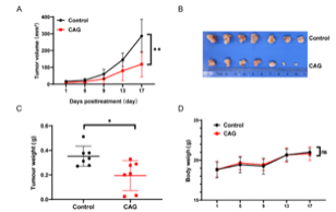
Figure 1: Changes in A) tumor volume, C) tumor weight, and D) body weight, with B) a visual representation of change in tumor size in treatment groups administered a vehicle vs CAG.
Furthermore, CAG effectively triggered both apoptosis and autophagy within the tumor microenvironment, as indicated by significant PARP cleavage and LC3 accumulation in tumor lysates from CAG-treated mice. The molecular analysis of tumor proteins in vivo also showed that CAG treatment led to a notable activation of p-AMPK and p-ULK1, and inactivation of p-mTOR, p-p70S6K, and p-4EBP1, mirroring the AMPK/ULK1/mTOR pathway modulation observed in in vitro studies. These in vivo results conclusively demonstrated that CAG exerts robust anti-tumor activity in NSCLC by inducing apoptosis and protective autophagy without causing systemic toxicity, thereby supporting its potential as a therapeutic agent for lung cancer. The study’s comprehensive findings highlight CAG’s dual mechanism of action and provide a strong rationale for exploring combination therapies that strategically target autophagy to enhance the efficacy of CAG in combating NSCLC [1].
2) The study completed by Feng et al revealed the neuroprotective mechanisms of cycloastragenol (CAG) in Parkinson’s disease (PD) models, demonstrating its capacity to mitigate neuroinflammation, oxidative stress, and neuronal damage through multifaceted cellular and molecular pathways. Initial proteomic analysis of striatum samples from PD mouse models provided crucial insights into CAG’s brain penetrance and its impact on protein expression. Liquid Chromatography-Mass Spectrometry (LC-MS) confirmed that intragastrically administered CAG effectively crossed the blood-brain barrier (BBB) and accumulated in brain tissue. Subsequent proteomic analysis revealed significant alterations in differentially expressed proteins (DEPs) across normal, model, and model+CAG groups, with 20 DEPs identified between modeling and normal, 12 between model+CAG and model, and 24 between model+CAG and normal. Histograms and heat maps further illustrated the intricate patterns of up- and down-regulated DEPs. Notably, CAG treatment was found to markedly influence cholesterol biosynthesis in PD mice, and Gene Ontology (GO) analysis indicated that DEPs were involved in regulating endoplasmic reticulum function [2].
A key finding from the proteomic data was the reduction of Scribble (Scrib) and eight other proteins in the model+CAG group. Colocalization analysis specifically confirmed that Scrib and Iba1, a microglial marker, were co-expressed, pointing to Scrib’s presence predominantly in microglia. In PD mice, both Scrib and p22phox levels were elevated, and these increases were significantly attenuated by CAG administration, suggesting that CAG plays a role in diminishing NADPH oxidase activity. Protein-Protein Interaction (PPI) networks, derived from the STRING database, further highlighted Scrib’s interaction with well-known PD-associated proteins like mitogen-activated protein kinase (MAPK) and leucine-rich-repeat kinase 2 (LRRK2). Crucially, immunofluorescence and Sholl’s analyses demonstrated that CAG suppressed NLRP3 inflammasome expression in microglia in vivo, without affecting astrocytes, and also mitigated aberrant microglial process branching and morphological changes observed in the PD model. These findings collectively underscored CAG’s ability to reduce Scrib/p22phox activity and NLRP3 inflammasome activation in PD mice.
Further in vitro investigations using primary microglial cultures revealed how CAG modulates microglial responses to α-synuclein (α-Syn) pathology. α-Syn pre-formed fibrils (PFFs) induced significant microglial activation, characterized by morphological changes and cell body enlargement. α-Syn stimulation also dramatically elevated Scrib and p22phox expression, leading to increased colocalization and reactive oxygen species (ROS) production in primary microglia. CAG treatment dose-dependently attenuated these α-Syn-induced increases in Scrib and p22phox expression, their colocalization, and subsequent ROS generation, suggesting a broad antioxidant effect by inhibiting NOX-mediated ROS generation [2].
CAG also suppressed α-Syn-induced CD68 upregulation (a marker of microglial activation and phagocytosis) in a dose-dependent manner and concurrently promoted α-Syn degradation. Moreover, α-Syn stimulation increased the mRNA levels of pro-inflammatory cytokines such as inducible nitric oxide synthase (iNOS), TNF-α, IL-1β, and IL-6, which were dose-dependently mitigated by CAG co-treatment. While α-Syn reduced anti-inflammatory markers like IL-10 and Ym1, CAG increased CD163 expression, indicating a reprogramming of microglia towards a more protective phenotype. These results strongly suggest that CAG reprograms microglia to reduce persistent α-Syn-triggered activation by enhancing phagocytosis and suppressing Scrib-associated ROS generation [2].
The study also delved into the impact of CAG on microglial autophagy and mitochondrial integrity. α-Syn stimulation led to an increase in p62 expression without affecting LC3 levels, indicating impaired autophagy. CAG effectively reduced this α-Syn-mediated p62 accumulation in a dose-dependent manner. However, co-administration with the autophagy inhibitor 3-MA hindered CAG’s ability to reduce p62, confirming its role in modulating autophagy. While α-Syn increased LC3-I, CAG enhanced LC3 lipididation, resulting in a higher LC3-II/LC3-I ratio, signifying improved autophagosome formation. TEM images showed that CAG maintained the ultrastructural integrity of microglia and alleviated α-Syn-induced mitochondrial damage. Intriguingly, CAG facilitated the engulfment of α-Syn into microglial autophagosomes, a crucial step for clearing toxic aggregates. Given that p62 mediates the autophagic degradation of damaged mitochondria, CAG diminished α-Syn-induced p62 accumulation on mitochondria (co-localized with Tom20), implying improved mitophagy. Furthermore, CAG treatment reduced α-Syn-induced mitochondrial ROS (mtROS) production, exerting antioxidant effects comparable to MitoQ, a targeted mitochondrial antioxidant. These findings solidify CAG’s antioxidant role by enhancing autophagy and protecting against mitochondrial damage-induced ROS.
Building on the link between ROS and NLRP3 inflammasome activation, the study demonstrated that CAG effectively suppressed α-Syn-induced NLRP3 inflammasome activation and pyroptosis in primary microglia. CAG dose-dependently inhibited both mRNA and protein expression of microglial NLRP3 inflammasomes, and reduced α-Syn-upregulated mature caspase-1 and cleaved gasdermin D (GSDMD), key components of the pyroptotic pathway. ELISA confirmed that CAG significantly inhibited the release of pro-inflammatory cytokines IL-1β and IL-18. Visualization of pyroptosis-linked pores via YO-PRO-1 iodide uptake showed that α-Syn-exposed microglia exhibited significant permeability, which was remarkably reduced by CAG, reversing GSDMD pore formation in a dose-dependent fashion [2].
Finally, CAG’s neuroprotective effects were investigated. Microglia-conditioned media (MCM) from α-Syn-activated microglia increased intracellular calcium levels in primary neurons, which was dose-dependently reduced by CAG-treated MCM. CAG-treated MCM also inhibited α-Syn-induced caspase-3 activation, apoptosis, nNOS upregulation, and nitrosative stress, in neurons. Sholl analysis revealed that CAG-treated MCM alleviated α-Syn-stimulated MCM-induced neuronal curling and reduced branching, while TEM images showed increased secondary lysosomes and preserved mitochondrial integrity in neurons exposed to CAG-treated MCM.
These results demonstrate that CAG mitigates microglia-mediated inflammation, normalizes intracellular Ca2+ levels, reduces nitrosative stress, and protects neurons from α-Syn-induced damage and apoptosis. In vivo behavioral assessments confirmed these neuroprotective effects: CAG significantly improved α-Syn overexpression-diminished motor coordination and balance (stride length, stance length, gait symmetry, beam walk, rotarod performance) and ameliorated anxiety-like behaviors. Furthermore, CAG treatment notably decreased α-Syn levels in the striatum and protected against the loss of TH-positive neurons in the substantia nigra pars compacta (SNpc), providing a holistic picture of CAG’s efficacy in ameliorating PD pathology [2].
Disclaimer
**LAB USE ONLY**
*This information is for educational purposes only and does not constitute medical advice. THE PRODUCTS DESCRIBED HEREIN ARE FOR RESEARCH USE ONLY. All clinical research must be conducted with oversight from the appropriate Institutional Review Board (IRB). All preclinical research must be conducted with oversight from the appropriate Institutional Animal Care and Use Committee (IACUC) following the guidelines of the Animal Welfare Act (AWA).
Citations
[1] Zhu LH, Liang YP, Yang L, Zhu F, Jia LJ, Li HG. Cycloastragenol induces apoptosis and protective autophagy through AMPK/ULK1/mTOR axis in human non-small cell lung cancer cell lines. J Integr Med. 2024;22(4):503-514. doi:10.1016/j.joim.2024.05.004
[2] Feng L, Lo H, Zheng J, Weng W, Sun Y, Pan X. Cycloastragenol reduces microglial NLRP3 inflammasome activation in Parkinson’s disease models by promoting autophagy and reducing Scrib-driven ROS. Phytomedicine. 2024;135:156210. doi:10.1016/j.phymed.2024.156210
Cycloastragenol is sold for laboratory research use only. Terms of sale apply. Not for human consumption, nor medical, veterinary, or household uses. Please familiarize yourself with our Terms & Conditions prior to ordering.
| File Name | View/Download |
| Coming Soon! |  |
VIEW CERTIFICATES OF ANALYSIS (COA)

Reviews
There are no reviews yet.