



Description
Myricetin Nootropic Powder (5 Grams)








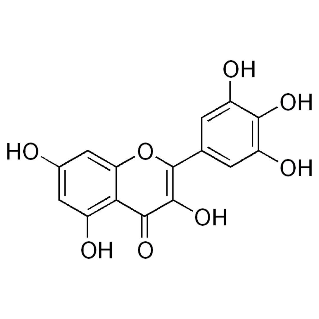
| CAS Number | 529-44-2 |
| Other Names | Cannabiscetin, Myricetol, Myricitin |
| IUPAC Name |
3,3′,4′,5,5′,7-Hexahydroxyflavone
|
| Molecular Formula | C₁₅H₁₀O₈ |
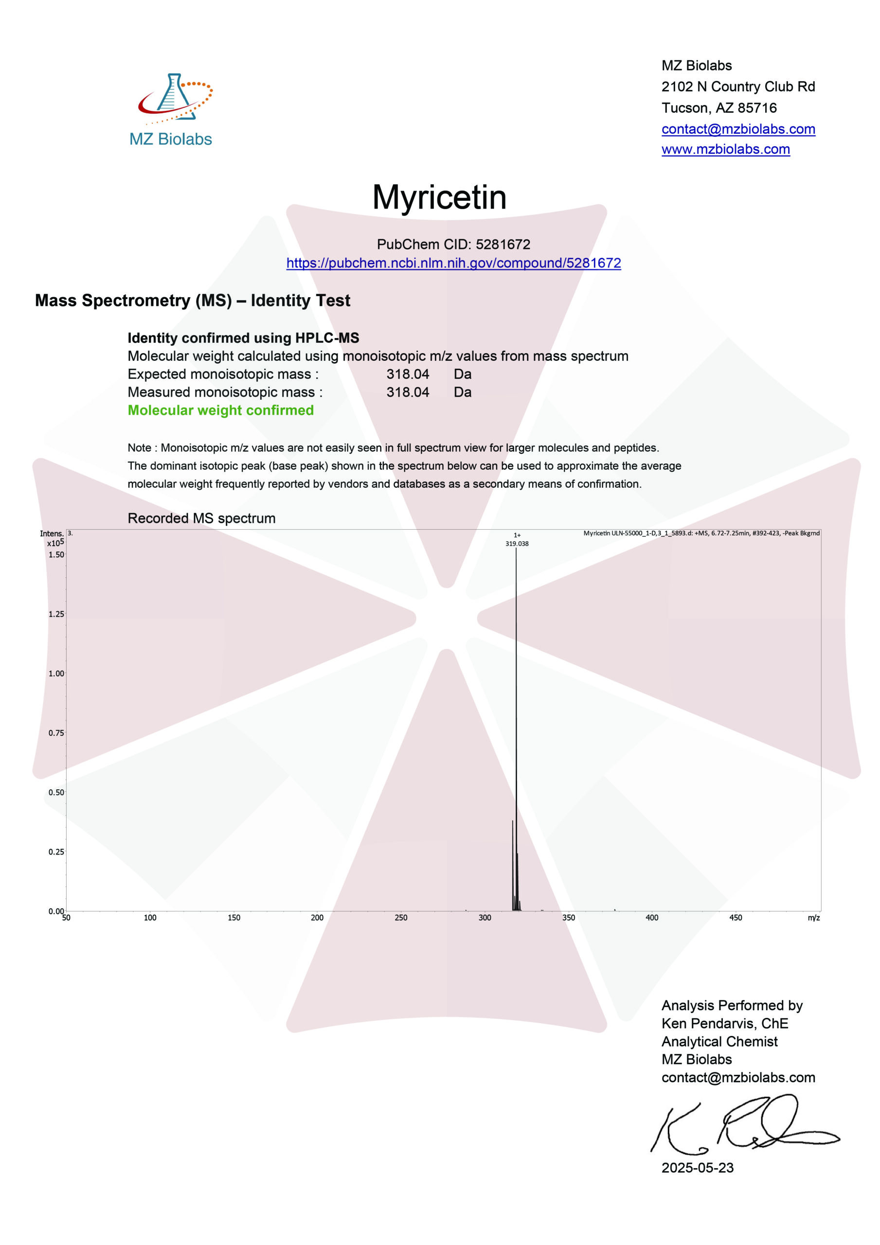
| Molecular Weight | 318.23 |
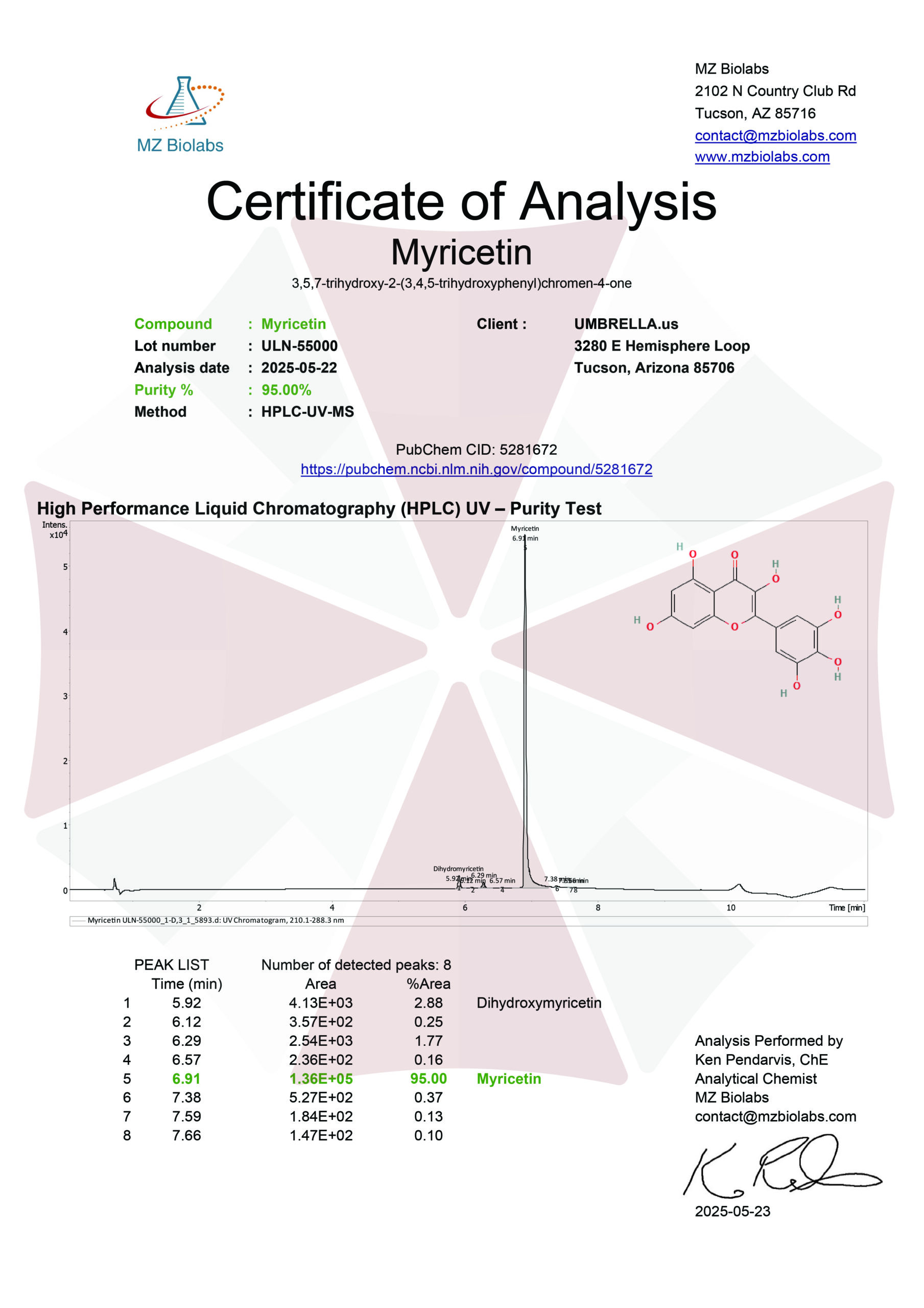
| Purity | ≥99% Pure (LC-MS) |
| Liquid Availability | N/A |
| Powder Availability |  5 grams |
| Gel Availability | N/A |
| Storage | Store in cool dry environment, away from direct sunlight. |
| Terms | All products are for laboratory developmental research USE ONLY. Products are not for human consumption. |
What is Myricetin?
Myricetin is classified among the polyphenolic compounds, and known for its potent antioxidant, anti-inflammatory, and neuroprotective properties. Myricetin has garnered increasing scientific interest as a potential nootropic agent as research suggests that the compound may support cognitive health by modulating oxidative stress, improving synaptic plasticity, and influencing signaling pathways involved in memory and learning, such as the PI3K/Akt and MAPK pathways. Preliminary studies have indicated its ability to cross the blood–brain barrier, further supporting its potential utility in enhancing brain function and possibly mitigating cognitive decline associated with neurodegenerative disorders like Alzheimer’s disease.
Main Research Findings
1) Myricetin was found to inhibit TNF-alpha-induced NF-KB activation in A549 cells resulting in reduced inflammatory responses.
2) Treatment with myricetin mitigated motor disturbances and decreased neuronal ferroptosis in animal models of Parkinson’s disease.
Selected Data
1) The study conducted by the team of Chen et al explored the biological effects of myricetin, a plant-derived flavonoid, on human lung adenocarcinoma cells (A549) using various cellular and molecular biology techniques. The A549 cells were cultured in DMEM and supplemented with 10% fetal bovine serum, at 37 °C in a humidified incubator with 5% CO₂. These conditions provided a stable environment to assess how myricetin affects cell viability, gene and protein expression, inflammatory signaling, and intracellular pathways [1].
To evaluate cytotoxicity and determine the appropriate myricetin concentrations for subsequent experiments, a CCK-8 assay was conducted. A549 cells were seeded into 96-well plates and treated with increasing concentrations of myricetin ranging from 7.8 µM to 1000 µM, for 24 hours. Following treatment, a CCK-8 reagent was added, and the optical density was measured at 450 nm using a microplate reader. This assay enabled the researchers to quantify the viability of cells post-treatment and identify concentrations that were non-toxic yet biologically effective [1].
To investigate the effects of myricetin on gene expression, total RNA was extracted from treated A549 cells and complementary DNA was synthesized from the extracted RNA. The resulting cDNA was then subjected to reverse transcription-quantitative polymerase chain reaction using SYBR Premix Ex Taq II to amplify target genes. The thermal cycling conditions included an initial denaturation at 95 °C for 2 minutes, followed by 40 amplification cycles of 95 °C for 10 seconds and 60 °C for 30 seconds. Specific primers used for gene amplification were listed in a corresponding table, enabling assessment of gene expression changes induced by myricetin.
Western blotting was performed to assess protein expression levels and post-translational modifications influenced by myricetin treatment. Proteins were extracted from A549 cells using a radioimmunoprecipitation assay buffer containing phenylmethylsulfonyl fluoride, followed by centrifugation at 12,000 rpm for 15 minutes at 4 °C. Protein concentrations were measured using a Bicinchoninic Acid assay. Equal protein amounts of 25 µg were separated using 10% SDS-PAGE and transferred to polyvinylidene difluoride membranes. After blocking with 5% non-fat milk, membranes were incubated with primary antibodies targeting key proteins in inflammatory and regulatory pathways, including IκB-α, total and phosphorylated NF-κB p65, SIRT1, acetyl-p65, acetyl-p53, total p65, p53, and loading controls. After overnight incubation at 4 °C, membranes were treated with horseradish peroxidase-conjugated secondary antibodies, and protein bands were visualized using a chemiluminescent substrate [1].
To further assess the inflammatory response, ELISA assays were performed to detect interleukin-6 (IL-6) and interleukin-8 (IL-8) levels in cell culture supernatants. A549 cells were first pretreated with myricetin, then stimulated with tumor necrosis factor-alpha (TNF-α) for 24 hours. After incubation, the culture supernatants were collected and centrifuged to remove cellular debris. Protein levels of IL-6 and IL-8 were quantified using ELISA kits according to the manufacturer’s protocol, allowing researchers to evaluate the anti-inflammatory potential of myricetin.
Immunofluorescence staining was employed to visualize the localization of key regulatory proteins. A549 cells were pretreated with various concentrations of myricetin at 20, 40, or 60 µM, or salermide, a SIRT1 inhibitor, at 10 µM, followed by TNF-α exposure for 24 hours. Cells were fixed with paraformaldehyde and permeabilized with Triton X-100. After blocking with bovine serum albumin, cells were incubated with antibodies against NF-κB p65 and SIRT1 overnight at 4 °C. FITC-conjugated secondary antibodies were used for detection, and nuclei were stained with DAPI. Stained cells were observed using a laser-scanning confocal microscope to assess the subcellular localization and activation status of the target proteins. This analysis helped determine whether myricetin influences the nuclear translocation of NF-κB p65 or modulates SIRT1 activity [1].
To directly measure SIRT1 enzymatic activity, a fluorescent activity assay was conducted. Nuclear proteins were extracted and processed using a SIRT1 Activity Assay Kit. The assay measured optical density at 405 nm, reflecting SIRT1’s deacetylase activity under different treatment conditions. This step was crucial in confirming whether myricetin’s regulatory effects were mediated through modulation of SIRT1 function [1].
In summary, this study systematically examined the influence of myricetin on lung adenocarcinoma A549 cells by integrating cytotoxicity testing, gene and protein expression analysis, cytokine measurement, immunofluorescence microscopy, and enzymatic activity assays. These comprehensive methodologies aimed to elucidate myricetin’s potential as an anti-inflammatory and anti-cancer agent, particularly through pathways involving SIRT1 and NF-κB signaling [1]
2) The research team of Gu et al completed a comprehensive experimental investigation into the protective effects of myricetin in a Parkinson’s disease (PD) model using both in vivo and in vitro systems. Various reagents, procedures, and assessments were used to elucidate the therapeutic potential of myricetin, with a focus on oxidative stress, ferroptosis, and neuroprotection [2].
The SH-SY5Y human neuroblastoma cell line was used for in vitro studies and cultured in standard DMEM supplemented with 10% fetal bovine serum under 5% CO₂ at 37°C. For treatment, the cells were exposed to 0.4 mM MPP⁺, a neurotoxin that mimics PD pathology, 50 μM myricetin, 1 μM erastin, as a ferroptosis inducer, or 1 μM ferrostatin-1, as a ferroptosis inhibitor, for 24 hours. The impact of these treatments was subsequently evaluated through various biochemical and molecular analyses.
A rat model of Parkinson’s disease was created using intraperitoneal injections of MPTP at a dose of 30 mg/kg/day for five days, a method known to induce dopaminergic neuron degeneration, particularly in the substantia nigra. Control rats received vehicle injections without MPTP. To test the therapeutic effect of myricetin, it was administered orally at 25 mg/kg/day from day 1 to day 14. In another experimental arm, erastin was delivered intranasally at 15 mg/kg immediately after MPTP injections. All rats were maintained under standard laboratory conditions with controlled temperature, humidity, and a 12-hour light/dark cycle. They had free access to food and water. Rats were weighed weekly from day 7, and euthanized after four weeks using an overdose of pentobarbital followed by cervical dislocation. Substantia nigra tissues were then harvested for further analysis [2].
Motor coordination and balance of the rats was assessed through behavioral tests including the rotarod, balance beam, and foot fault tests. In the rotarod test, rats were tested on their ability to stay on a rotating rod that accelerated from 4 to 40 rpm over five minutes. The time before falling was recorded across three trials. The balance beam test evaluated rats’ ability to walk across a narrow wooden beam elevated 60 cm above the ground. Rats were scored from 1 to 6 based on performance, ranging from falling immediately to walking without any issues. The foot fault test involved a mesh platform where rats’ foot slips were counted during a 1-minute ambulation period. Foot fault percentage was calculated by the ratio of missteps to total steps, multiplied by 100% [2].
Western blotting was employed to assess protein expression levels in both substantia nigra tissue and SH-SY5Y cells. Proteins were extracted using a RIPA buffer, and concentrations were measured using a Pierce BCA assay. (20 μg of proteins per sample were separated by SDS-PAGE, transferred to PVDF membranes, blocked with 5% non-fat milk, and incubated with specific primary antibodies overnight at 4°C. This was followed by secondary antibody incubation and signal detection using ChemiDox XRS imaging and ImageJ analysis, with β-actin and Lamin B1 serving as loading controls.
Immunohistochemical staining of substantia nigra tissues was performed to localize specific proteins. Fixed tissues were paraffin-embedded, sectioned, and processed through standard dewaxing, hydration, antigen retrieval using citrate buffer, and blocking steps. Primary antibodies were applied, followed by HRP-conjugated secondary antibodies. Visualization was achieved using an HRP DAB kit, and counterstaining was done with hematoxylin [2].
Reactive oxygen species levels were measured in both substantia nigra tissues and SH-SY5Y cells using the DCFDA fluorescent probe. Substantia nigra tissue was homogenized, incubated with DCFDA for 30 minutes, and fluorescence intensity was measured with a microplate reader. SH-SY5Y cells were similarly treated with DCFDA, fixed with paraformaldehyde, and fluorescence was visualized under a microscope and quantified using ImageJ software.
Iron content was determined using an iron assay kit. Substantia nigra tissue and SH-SY5Y cells were homogenized in an assay buffer, centrifuged, and supernatants were incubated with an iron reducer and probe, while absorbance was measured at 593 nm. This assay helps assess ferroptosis, as iron accumulation is a hallmark of this form of cell death. Lipid peroxidation, a sign of oxidative stress, was evaluated by measuring malondialdehyde levels using an MDA assay kit. Absorbance was read at 532 nm. Glutathione levels, representing antioxidant capacity, were measured using a total glutathione assay kit and read at 412 nm [2].
Statistical analyses were performed using the Student’s t-test for two-group comparisons and one-way ANOVA with Scheffé post hoc tests for multiple comparisons. All experiments were conducted in triplicate, and results were expressed as mean ± standard deviation. In summary, this study employs a set of biochemical, molecular, and behavioral assessments to evaluate the neuroprotective effects of myricetin in PD models. By using both in vitro and in vivo systems and focusing on markers of oxidative stress, ferroptosis, and neuronal integrity, it provides insights into myricetin’s therapeutic potential [2].
Discussion
1) The study completed by the research team of Chen et al investigates the anti-inflammatory effects and molecular mechanisms of myricetin in TNF-α-stimulated A549 lung epithelial cells, which serve as a model for airway inflammation. The research focuses on how myricetin modulates pro-inflammatory cytokine expression and signaling pathways, particularly through the NF-κB and SIRT1 axes [1].
To determine a safe and effective concentration of myricetin for experiments, cytotoxicity assays were conducted using the CCK-8. A549 cells were treated with increasing concentrations of myricetin, ranging from 0 to 1000 μM, over a 24-hour period. Results showed that cell viability remained unaffected at concentrations up to 250 μM. Based on this observation, the researchers selected 20, 40, and 60 μM concentrations of myricetin for the subsequent experiments, ensuring therapeutic efficacy without cytotoxic effects.
One of the key pro-inflammatory stimuli in inflammatory diseases is TNF-α, which upregulates cytokines such as IL-6 and IL-8. In the study, A549 cells stimulated with TNF-α showed a significant increase in the mRNA levels of both IL-6 and IL-8. However, treatment with myricetin effectively reduced the expression of these pro-inflammatory cytokines at both the mRNA and protein levels. This downregulation was also observed in human bronchial epithelial cells, suggesting that the anti-inflammatory effects of myricetin are not limited to a single cell type. These findings establish myricetin as a potential anti-inflammatory agent that suppresses key cytokines involved in airway inflammation [1].
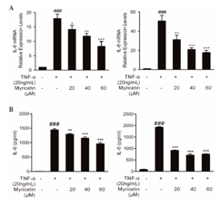
Figure 1: Changes in the expression of IL-6 and IL-8 levels expressed in A) mRNA of A549 cells and B) protein concentrations in the culture supernatant.
Since NF-κB is a central transcriptional regulator in inflammation, particularly for IL-6 and IL-8, the study further explored whether myricetin exerts its anti-inflammatory effects by interfering with the NF-κB signaling pathway. TNF-α stimulation was found to trigger degradation of IκB-α, a key inhibitor of NF-κB, and promote phosphorylation and nuclear translocation of the NF-κB p65 subunit in A549 cells. Pretreatment with myricetin significantly reversed these effects, indicating that myricetin inhibits activation of the NF-κB pathway. Immunofluorescence imaging confirmed that myricetin suppressed TNF-α-induced nuclear translocation of NF-κB p65, further validating its inhibitory effect on this inflammatory signaling cascade. Consistent results were observed in HBE cells, reinforcing the reliability of these findings [1].
Given the known role of SIRT1, a NAD+-dependent deacetylase, in regulating NF-κB activity, the researchers investigated whether SIRT1 was involved in the myricetin-mediated anti-inflammatory response. TNF-α was shown to increase acetylation of the NF-κB p65 subunit, a modification that enhances its transcriptional activity. Myricetin treatment reduced this acetylation, suggesting enhanced deacetylase activity, potentially through SIRT1. Interestingly, myricetin did not significantly change SIRT1 mRNA or total protein expression levels, nor its subcellular localization. However, it did reduce phosphorylation of SIRT1 at Ser47, a modification associated with its inhibition, implying a functional activation of SIRT1 in the presence of myricetin [1].
To validate the mechanistic link between SIRT1 activation and NF-κB inhibition, blocking experiments were performed using salermide, a specific SIRT1 antagonist. Salermide effectively reversed the myricetin-mediated reduction in IL-6 and IL-8 expression, both at the mRNA and protein levels. Furthermore, it negated the inhibitory effects of myricetin on NF-κB p65 phosphorylation, acetylation, and nuclear translocation, as well as the degradation of IκB-α. Salermide also reversed the myricetin-induced decrease in acetyl-p53 levels, confirming its role in suppressing SIRT1 activity. Importantly, while salermide did not affect SIRT1 expression levels, it significantly impaired its functional activity, thereby nullifying myricetin’s anti-inflammatory effects.
Collectively, these results demonstrate that myricetin exerts its anti-inflammatory effects by enhancing the activity of SIRT1, which in turn inhibits NF-κB signaling. This leads to decreased expression of pro-inflammatory cytokines IL-6 and IL-8 in TNF-α-stimulated A549 cells. The study presents a detailed mechanistic pathway in which myricetin acts not only as an inhibitor of NF-κB signaling but also as a modulator of SIRT1 activity, emphasizing its therapeutic potential for managing inflammatory diseases, particularly those involving airway inflammation [1].
In summary, this investigation provides evidence that myricetin is a potent anti-inflammatory compound capable of modulating key signaling pathways involved in cytokine production. Through activation of SIRT1 and subsequent suppression of NF-κB signaling, myricetin effectively reduces inflammation in airway epithelial cells, offering promising therapeutic avenues for treating inflammatory respiratory disorders [1].
2) This study conducted by Gu et al explores the neuroprotective effects of myricetin in PD using both in vivo and in vitro models. Specifically, the research focuses on how myricetin mitigates motor dysfunction, dopaminergic neuron loss, α-synuclein accumulation, and ferroptosis, a form of regulated cell death associated with iron overload and oxidative stress. The mechanistic pathway investigated includes the activation of the Nrf2/Gpx4 signaling axis, known for its role in counteracting ferroptosis [2].
To model PD in rats, MPTP was administered intraperitoneally to induce dopaminergic neuron degeneration. Rats were then treated orally with myricetin. MPTP treatment led to a significant reduction in body weight gain, indicating systemic and neurological stress. However, myricetin administration effectively restored normal weight gain patterns, suggesting overall health improvement. Behavioral tests including the beam balance test, foot fault test, and rotarod test were used to evaluate motor coordination and function. MPTP-treated rats exhibited impaired motor behavior across all tests, while myricetin significantly improved performance. Importantly, erastin, a specific ferroptosis inducer, partially reversed the motor improvements induced by myricetin in the foot fault test, though it had no significant effect in the beam balance and rotarod tests, indicating a partial dependence on ferroptosis regulation [2].
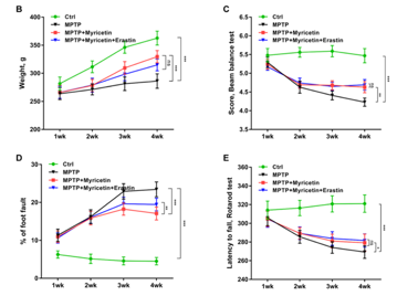
Figure 2: Changes in B) weight in grams, C) balance beam test score, D) percent of foot fault, and E) latency to fall assessed through the rotarod test, throughout the control group, the MPTP group, the MPTP + myricetin group, and the MPTP + myricetin + erastin group
To determine if myricetin could protect dopaminergic neurons, the study evaluated tyrosine hydroxylase, a marker of dopamine neurons, and α-synuclein, a key pathological protein in PD. Western blotting and immunohistochemistry of the substantia nigra showed that MPTP dramatically increased α-synuclein accumulation and reduced tyrosine hydroxylase expression, signifying neuronal loss. Myricetin reversed these changes, decreasing α-synuclein levels and restoring tyrosine hydroxylase protein expression. Furthermore, immunohistochemistry analysis confirmed an increase in tyrosine hydroxylase-positive cells in the substantia nigra following myricetin treatment. Again, these beneficial effects were inhibited by erastin, further indicating the involvement of ferroptosis in MPTP-induced neurotoxicity and myricetin’s protective effects.
Given that ferroptosis is characterized by iron overload and oxidative stress, the study assessed iron, reactive oxygen species, malondialdehyde, and glutathione levels in both substantia nigra tissue and serum. MPTP significantly elevated iron and reactive oxygen species levels, along with an increase in malondialdehyde, a marker of lipid peroxidation, and a decrease in GSH, an essential antioxidant. Myricetin treatment reversed all these changes, strongly indicating that it alleviates ferroptosis. When co-administered with erastin, these effects were blocked, confirming that ferroptosis inhibition is a critical mechanism of myricetin’s neuroprotection [2].
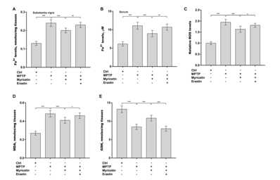
Figure 3: Changes in A) iron levels in the substantia nigra, B) iron levels in the serum, C) relative reactive oxygen species levels, D) malodialdehyde levels in the tissues, and E) GSH levels in the tissues
To understand whether motor function improvements correlated with ferroptosis inhibition, the researchers analyzed relationships between latency to fall assessed through the rotarod test, and ferroptosis biomarkers. No significant correlation was found between latency to fall and iron in the substantia nigra; however, serum iron and substantia nigra malondialdehyde levels showed a negative correlation with motor performance, while GSH levels had a positive correlation, supporting the hypothesis that oxidative stress and iron metabolism are tightly linked to motor dysfunction in PD [2].
The effects of myricetin were further validated in in vitro experiments using SH-SY5Y cells, a human neuroblastoma cell line commonly used to model PD. Exposure to MPP⁺, a toxic metabolite of MPTP, mimicked PD-like pathology by reducing cell viability and inducing ferroptosis. Treatment with Fer-1, a ferroptosis inhibitor, or myricetin restored cell viability. Myricetin also reduced iron, reactive oxygen species, and malondialdehyde levels while increasing GSH in MPP⁺-treated cells. Once again, these effects were nullified by co-treatment with erastin, reinforcing the role of ferroptosis inhibition in myricetin’s protective mechanism.
A crucial part of this study was the investigation of the Nrf2 (nuclear factor erythroid 2–related factor 2) pathway and its downstream effector Gpx4 (glutathione peroxidase 4), both of which are known to suppress ferroptosis. In MPP⁺-treated SH-SY5Y cells, total Nrf2 protein levels were not significantly altered. However, treatment with myricetin increased both total and nuclear Nrf2 levels, indicating enhanced activation and translocation of the transcription factor. Furthermore, MPP⁺ treatment caused a decrease in Gpx4 protein expression, which was reversed by myricetin, suggesting that the protective effects are mediated by Nrf2-driven upregulation of Gpx4 [2].
In conclusion, the study demonstrates that myricetin exerts strong neuroprotective effects in both in vivo and in vitro PD models. It improves motor function, protects dopaminergic neurons, and reduces α-synuclein accumulation. These effects are mechanistically linked to suppression of ferroptosis, evidenced by reduced iron and oxidative stress markers, and are mediated by activation of the Nrf2/Gpx4 pathway. The partial reversal of myricetin’s benefits by erastin underscores the critical role of ferroptosis inhibition. Taken together, these findings suggest that myricetin may be a promising candidate for therapeutic development in Parkinson’s disease and potentially other neurodegenerative conditions involving ferroptosis and oxidative stress [2].
Disclaimer
**LAB USE ONLY**
*This information is for educational purposes only and does not constitute medical advice. THE PRODUCTS DESCRIBED HEREIN ARE FOR RESEARCH USE ONLY. All clinical research must be conducted with oversight from the appropriate Institutional Review Board (IRB). All preclinical research must be conducted with oversight from the appropriate Institutional Animal Care and Use Committee (IACUC) following the guidelines of the Animal Welfare Act (AWA).
Citations
[1] Chen M, Chen Z, Huang D, et al. Myricetin inhibits TNF-α-induced inflammation in A549 cells via the SIRT1/NF-κB pathway [published correction appears in Pulm Pharmacol Ther. 2021 Jun;68:102031. doi: 10.1016/j.pupt.2021.102031.]. Pulm Pharmacol Ther. 2020;65:102000. doi:10.1016/j.pupt.2021.102000.
[2] Gu SC, Xie ZG, Gu MJ, et al. Myricetin mitigates motor disturbance and decreases neuronal ferroptosis in a rat model of Parkinson’s disease. Sci Rep. 2024;14(1):15107. Published 2024 Jul 2. doi:10.1038/s41598-024-62910-6
Myricetin is sold for laboratory research use only. Terms of sale apply. Not for human consumption, nor medical, veterinary, or household uses. Please familiarize yourself with our Terms & Conditions prior to ordering.


| File Name | View/Download |
| 2025-05-22-Umbrella-Labs-Myricetin-Certificate-of-Analysis-COA.pdf |  |
VIEW CERTIFICATES OF ANALYSIS (COA)


Reviews
There are no reviews yet.