




Description
Nootkatone Nootropic Powder






| CAS Number | 4674-50-4 |
| Other Names | (+)-Nootkatone, (+/-)-Nootkatone, Nootkanone, UNII-3K3OKV2A5A, 3K3OKV2A5A, IZ2Y119N4J, DTXSID8047050 |
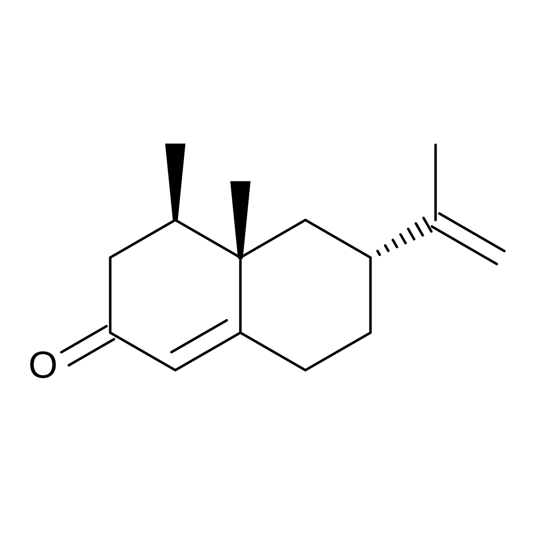
| IUPAC Name |
4α,5α-Eremophila-1(10),11-dien-2-one
|
| Molecular Formula | C₁₅H₂₂O |
| Molecular Weight | 218.34 |
| Purity | ≥99% Pure (LC-MS) |
| Liquid Availability | N/A |
| Powder Availability |  25 grams, 10 grams, 5 grams |
| Gel Availability | N/A |
| Storage | Store in cool dry environment, away from direct sunlight. |
| Terms | All products are for laboratory developmental research USE ONLY. Products are not for human consumption. |
What is Nootkatone?
Nootkatone is a naturally occurring compound best known for giving grapefruit its distinctive aroma, but in recent years it has attracted growing attention for its potential as a nootropic. Found in the essential oils of citrus fruits and certain plants like Alaska yellow cedar, nootkatone belongs to a class of organic compounds called sesquiterpenes. Beyond its use in flavoring and fragrances, emerging research suggests that nootkatone may influence energy metabolism, mitochondrial function, and cellular signaling pathways in ways that could support cognitive performance and brain health. As interest in plant-derived cognitive enhancers expands, nootkatone stands out as a promising candidate warranting further investigation.
Main Research Findings
1) Treatment with Nootkatone was found to reduce alveolar macrophage activation to protect against LPS-induced acute lung injury
2) Supplementation with Nootkatone was found to reduce nephrotoxicity induced through exposure of carbon tetrachloride.
Selected Data
1) The study completed by the research team of Cui et al employed a comprehensive in vivo and ex vivo experimental design to investigate the anti-inflammatory and immunometabolic effects of nootkatone in a lipopolysaccharide (LPS)-induced acute lung injury (ALI) mouse model. Female C57BL/6N mice, aged 6–8 weeks and weighing 18–22 grams, were maintained under pathogen-free conditions and randomly divided into five experimental groups: a normal saline (NS) control group, an LPS-induced ALI group, and three treatment groups receiving LPS combined with different doses of nootkatone ranging between 20, 40, and 80 mg/kg. ALI was induced via intratracheal instillation of LPS at a dose of 5 mg/kg, while nootkatone was administered orally one hour prior to LPS exposure. Mice were euthanized 24 hours after induction to allow for the assessment of inflammatory responses, lung pathology, and cellular metabolism [1].
Bronchoalveolar lavage fluid (BALF) was collected through tracheal cannulation and repeated phosphate-buffered saline washes. This fluid was used to quantify immune cell infiltration and cytokine production. Total cell counts were determined using a hemocytometer, while flow cytometry enabled identification of specific immune populations, including leukocytes, neutrophils, and alveolar macrophages (AMs), based on cell surface markers such as CD45, CD11c, Siglec-F, and Gr1. Additional flow cytometric analysis distinguished macrophage polarization states by measuring markers associated with pro-inflammatory (M1) and anti-inflammatory (M2) phenotypes, including CD80 and Arg1. These approaches allowed for precise characterization of immune cell dynamics within the lung microenvironment [1].
Histopathological analysis was conducted on lung tissue sections stained with hematoxylin and eosin. Tissue damage was evaluated using a standardized lung injury scoring system that assessed neutrophil infiltration, interstitial inflammation, edema, and vascular congestion. Each parameter was graded on a scale from 0 to 4, and total scores were calculated by summing individual categories. Importantly, scoring was performed by a blinded pathologist to reduce bias. To further investigate cellular mechanisms, primary alveolar macrophages were isolated from lung tissue באמצעות enzymatic digestion followed by magnetic separation using Siglec-F-specific microbeads, ensuring a highly purified macrophage population for downstream analyses.
Cytokine levels in BALF, specifically IL-6, TNF-α, and IL-1β, were quantified using enzyme-linked immunosorbent assays, providing a measure of inflammatory signaling. In parallel, cellular metabolism was assessed using Seahorse extracellular flux analysis. Glycolytic activity was measured via proton efflux rate (PER), while mitochondrial respiration was evaluated through oxygen consumption rate (OCR). These assays enabled detailed characterization of metabolic pathways, including basal respiration, ATP production, maximal respiration, glycolysis, and compensatory glycolysis. Pharmacological inhibitors such as rotenone/antimycin A and 2-deoxyglucose were applied during these assays to dissect metabolic contributions from mitochondrial and glycolytic processes.
At the molecular level, protein expression and signaling pathway activation were analyzed using immunoblotting. Lung tissues and isolated AMs were lysed, and proteins were separated by SDS-PAGE before being probed with antibodies targeting both total and phosphorylated forms of key signaling proteins, including STING, TBK1, and IRF3. These analyses allowed the researchers to evaluate activation of the STING/TBK1/IRF3 pathway, a central regulator of innate immune responses. Gene expression profiling was further explored באמצעות bulk RNA sequencing. RNA extracted from lung tissues and AMs underwent library preparation and sequencing on an Illumina platform. Transcript levels were normalized and visualized באמצעות heatmaps, enabling identification of differentially expressed genes related to inflammation and metabolism [1].
Additional assays were conducted to examine oxidative stress and molecular interactions. Mitochondrial reactive oxygen species (ROS) production in AMs was visualized באמצעות MitoSOX staining and confocal microscopy. Bioinformatics tools such as SwissTargetPrediction and WikiPathways were used to predict potential molecular targets of nootkatone and identify enriched signaling pathways. Molecular docking simulations were performed to model interactions between nootkatone and candidate proteins, particularly AKT1, providing mechanistic insight into how nootkatone may influence intracellular signaling. Finally, statistical analyses were conducted using one-way ANOVA followed by Dunnett’s post hoc test, with significance defined as p < 0.05, ensuring rigorous evaluation of experimental differences across groups [1].
2) The study by Dai et al. investigated the nephroprotective effects of nootkatone (NKT) against carbon tetrachloride (CCl₄)-induced kidney injury in mice using a controlled experimental design. The researchers employed a combination of biochemical assays, histopathological analysis, molecular biology techniques, and immunohistochemistry to comprehensively evaluate kidney function, oxidative stress, inflammation, and apoptosis.
Nootkatone, with a purity greater than 97%, was obtained commercially and prepared as a suspension in 0.5% sodium carboxymethyl cellulose (CMC-Na) at concentrations of 0.5, 1, and 2 mg/mL. Carbon tetrachloride, the nephrotoxic agent used to induce acute kidney injury, was also commercially sourced. Various biochemical assay kits were used to measure oxidative stress markers and enzyme activities, including catalase (CAT), superoxide dismutase (SOD), glutathione (GSH), glutathione peroxidase (GPX), inducible nitric oxide synthase (iNOS), and malondialdehyde (MDA). Enzyme-linked immunosorbent assay kits were used to quantify inflammatory cytokines such as TNF-α, IL-1β, and IL-6 [2].
The in vivo experiment utilized forty-eight male C57BL/6 mice aged eight weeks and weighing approximately 20–22 grams. The animals were housed under controlled environmental conditions, including a temperature of around 25°C, 55% humidity, and a 12-hour light/dark cycle, with free access to food and water. The mice were randomly divided into six groups of eight animals each: a control group, a CCl₄-treated group, three groups receiving different doses of NKT ranging between 5, 10, and 20 mg/kg/day, followed by CCl₄ exposure, and a group receiving NKT alone.
NKT was administered orally once daily for seven consecutive days before CCl₄ exposure. Acute kidney injury was induced by administering CCl₄, and the mice were euthanized 24 hours later using intraperitoneal injection of pentobarbital sodium. Blood samples were collected for serum analysis, and kidney tissues were harvested for histological, biochemical, and molecular analyses. One portion of the kidney was fixed in formaldehyde for histopathological examination, while the remaining tissue was stored at −80°C for protein and gene expression studies [2].
Kidney function was assessed by measuring serum levels of blood urea nitrogen (BUN) and creatinine (CRE) using an automated biochemical analyzer. Histopathological evaluation involved fixing kidney tissues, sectioning, staining, and scoring tissue damage using a semi-quantitative scoring system that assessed structural abnormalities such as tubular degeneration, necrosis, and glomerular damage. To evaluate oxidative stress, kidney tissues were homogenized and centrifuged to obtain supernatants. These were analyzed for levels of MDA, GSH, and nitric oxide (NO), as well as activities of antioxidant enzymes CAT, SOD, GPX, and iNOS. All measurements were normalized to total protein concentration determined by a BCA protein assay. These assays allowed for quantification of both oxidative damage and antioxidant defense capacity [2].
Inflammatory responses were assessed by measuring protein levels of IL-1β, IL-6, and TNF-α in kidney tissue using ELISA kits. Apoptotic activity was evaluated by measuring the activities of caspase-3 and caspase-9, key enzymes involved in the mitochondrial apoptosis pathway. Tissue lysates were prepared and analyzed using commercial activity kits, with results normalized to protein content. Immunohistochemistry was used to assess the expression of NOX4, an enzyme involved in reactive oxygen species production. Kidney tissue sections were stained using specific antibodies, and staining intensity was evaluated using a semi-quantitative scoring system based on color intensity across multiple microscopic fields.
Gene expression analysis was performed using quantitative reverse transcription polymerase chain reaction (qRT-PCR). Total RNA was extracted from kidney tissues, converted into cDNA, and amplified using specific primers for genes involved in oxidative stress and inflammation, including NF-κB, IL-1β, IL-6, TNF-α, iNOS, NOX4, Nrf2, and HO-1. β-actin served as the internal control, and relative gene expression was calculated. This comprehensive methodological approach enabled the researchers to evaluate the protective effects of NKT across multiple biological levels, including functional, structural, biochemical, and molecular endpoints [2].
Discussion
1) The results of the study by Cui et al demonstrated that nootkatone exerts significant protective effects against LPS-induced acute lung injury by reducing inflammation, modulating immune cell behavior, and reprogramming cellular metabolism. In the ALI mouse model, LPS exposure led to a marked increase in total cell counts, macrophages, and neutrophils in BALF, indicating robust immune cell infiltration into the lungs. Treatment with nootkatone significantly reduced these cell populations in a dose-dependent manner, with the highest dose of 80 mg/kg producing the most pronounced effect. Additionally, levels of key pro-inflammatory cytokines, IL-6, TNF-α, and IL-1β, were substantially elevated following LPS administration but were significantly suppressed by nootkatone treatment. Histological examination further confirmed these findings, showing that nootkatone alleviated lung tissue damage, including edema, inflammatory infiltration, and structural disruption [1].
Beyond its anti-inflammatory effects, nootkatone markedly influenced metabolic processes within lung tissue. LPS exposure increased mitochondrial respiration, as indicated by elevated OCR and associated parameters such as basal respiration, maximal respiration, ATP production, and spare respiratory capacity. Nootkatone treatment reversed these increases, bringing metabolic activity closer to baseline levels observed in control mice. Similarly, glycolytic activity, measure PER, was significantly elevated in LPS-treated mice, reflecting a shift toward high-energy metabolic states typical of inflammation. Nootkatone reduced glycolysis, including basal and compensatory glycolytic rates, suggesting that it restores metabolic balance in inflamed lung tissue.
At the molecular signaling level, the study identified the STING/TBK1/IRF3 pathway as a key mediator of inflammation in ALI. LPS stimulation increased phosphorylation of STING, TBK1, and IRF3, indicating activation of this pathway. Nootkatone significantly reduced phosphorylation of these proteins without affecting their total expression levels, demonstrating selective inhibition of pathway activation. This suppression was accompanied by decreased expression of IRF3-regulated inflammatory genes, including chemokines and cytokines involved in immune cell recruitment and activation. These findings suggest that nootkatone mitigates inflammation by targeting upstream signaling mechanisms that regulate cytokine production [1].
The study also provided detailed insights into the role of AMs, which are central to lung immune responses. In AMs isolated from LPS-treated mice, both mitochondrial respiration and glycolysis were significantly increased, mirroring the metabolic changes observed in whole lung tissue. Nootkatone reversed these metabolic alterations, reducing OCR and PER parameters and lowering mitochondrial ROS production. RNA sequencing data supported these findings, showing that genes associated with metabolic pathways were upregulated in LPS conditions and normalized following nootkatone treatment. This indicates that nootkatone not only suppresses inflammation but also restores normal metabolic function in immune cells [1].
In terms of immune phenotype, LPS disrupted macrophage homeostasis by increasing expression of both M1, pro-inflammatory. and certain M2, anti-inflammatory markers, reflecting a dysregulated and mixed activation state. Nootkatone treatment rebalanced macrophage polarization, reducing excessive expression of inflammatory markers while restoring key M2-associated markers such as CD206 and Ym1. Flow cytometry confirmed these changes, demonstrating normalization of CD80 and Arg1 expression levels. This suggests that nootkatone promotes a return to a more stable and functional immune phenotype rather than simply suppressing immune activity.
Finally, mechanistic analyses identified AKT1 as a potential molecular target of nootkatone. Computational docking studies showed that nootkatone binds effectively to AKT1, potentially interfering with its catalytic activity. Given that AKT1 is known to regulate the STING pathway, this interaction provides a plausible explanation for the observed inhibition of STING/TBK1/IRF3 signaling. Overall, the results demonstrate that nootkatone protects against acute lung injury through a multifaceted mechanism involving suppression of inflammatory signaling, normalization of immune cell metabolism, and restoration of macrophage function. These findings position nootkatone as a promising candidate for further therapeutic development in inflammatory lung diseases [1].
2) The results of the study performed by Dai et al demonstrated that NKT exerts significant protective effects against carbon tetrachloride (CCl₄)-induced nephrotoxicity in mice through modulation of oxidative stress, inflammation, apoptosis, and key molecular signaling pathways. These findings were supported by consistent improvements across biochemical, histological, and molecular indicators of kidney injury [2].
First, kidney function was evaluated by measuring serum levels of BUN and CRE, both of which are standard biomarkers of renal impairment. CCl₄ exposure caused a marked increase in BUN and CRE levels compared to the control group, confirming successful induction of acute kidney injury. However, pretreatment with NKT significantly reduced these elevated levels in a dose-dependent manner. The highest dose of NKT of 20 mg/kg/day produced the most pronounced improvement, bringing BUN and CRE values closer to normal levels. Importantly, NKT alone did not affect these parameters, indicating that it does not adversely impact kidney function under normal conditions.
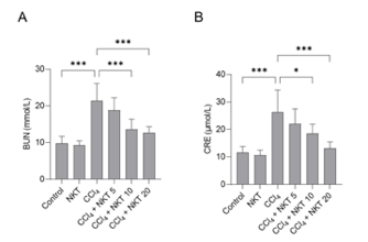
Figure 1: Changes in levels of A) BUN and B) CRE across experimental treatment groups.
Histopathological analysis further supported these findings. Kidney tissues from CCl₄-treated mice exhibited severe structural damage, including tubular degeneration, necrosis, dilation, cast formation, and glomerular congestion and hemorrhage. In contrast, mice pretreated with NKT showed substantial attenuation of these pathological changes. The degree of tissue damage, quantified using a semi-quantitative scoring system, was significantly reduced in the NKT-treated groups, particularly at higher doses. These observations demonstrate that NKT preserves renal structural integrity in the face of toxic insult [2].
Oxidative stress markers revealed another key aspect of NKT’s protective mechanism. CCl₄ exposure significantly increased levels of MDA and NO, both indicators of oxidative damage, while simultaneously reducing levels of glutathione (GSH) and activities of antioxidant enzymes such as SOD, CAT, and GPX. NKT pretreatment effectively reversed these changes. Specifically, it reduced MDA and NO levels while restoring GSH content and enhancing antioxidant enzyme activities. These effects were dose-dependent, with higher doses of NKT producing greater improvements. This indicates that NKT enhances the endogenous antioxidant defense system and mitigates lipid peroxidation and oxidative injury.
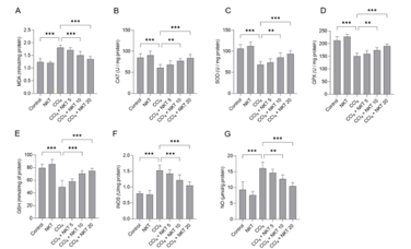
Figure 2: Changes in A) MDA, B) CAT, C) SOD, D) GPX, E) GSH, F) iNOS, and G) NO across experimental treatment groups.
Inflammatory responses were also significantly modulated by NKT. CCl₄ exposure led to marked increases in pro-inflammatory cytokines IL-1β, IL-6, and TNF-α in kidney tissues. NKT pretreatment significantly reduced the levels of these cytokines, again in a dose-dependent manner. At the highest dose, cytokine levels were substantially lowered, indicating strong anti-inflammatory effects. NKT alone did not alter cytokine levels, suggesting that its anti-inflammatory action is specifically relevant under pathological conditions [2].
Apoptosis, a critical contributor to tissue injury, was assessed by measuring the activities of caspase-3 and caspase-9. CCl₄ treatment significantly increased the activities of both enzymes, indicating activation of the mitochondrial apoptotic pathway. NKT pretreatment significantly suppressed this activation, reducing caspase activity in a dose-dependent fashion. These results suggest that NKT protects kidney cells from programmed cell death induced by toxic stress. At the molecular level, the study examined expression of proteins and genes involved in oxidative stress and inflammation. Immunohistochemical analysis showed that CCl₄ markedly increased expression of NOX4, a key enzyme involved in reactive oxygen species generation. NKT pretreatment significantly reduced NOX4 expression, indicating suppression of a major source of oxidative stress [2].
Gene expression analysis using qRT-PCR revealed that CCl₄ upregulated pro-inflammatory and stress-related genes, including NF-κB, IL-1β, IL-6, TNF-α, iNOS, and NOX4. NKT pretreatment significantly downregulated these genes, demonstrating inhibition of inflammatory and oxidative signaling pathways. Conversely, NKT upregulated the expression of Nrf2 and HO-1, which are central components of the antioxidant defense system. Activation of the Nrf2/HO-1 pathway is known to enhance cellular resistance to oxidative stress, and its upregulation provides a mechanistic explanation for the observed protective effects.
Notably, these molecular changes were dose-dependent and most pronounced at the highest NKT concentration. Additionally, NKT alone produced mild beneficial effects on gene expression without causing harmful changes, further supporting its safety profile. Overall, the results indicate that NKT protects against CCl₄-induced kidney injury through multiple interconnected mechanisms. These include reducing oxidative stress by inhibiting NOX4 and enhancing antioxidant defenses, suppressing inflammation via downregulation of NF-κB and cytokines, and preventing apoptosis through inhibition of caspase activation. The simultaneous activation of the Nrf2/HO-1 pathway further strengthens the cellular defense system. In conclusion, the findings provide strong evidence that nootkatone has potent nephroprotective effects in this experimental model. By targeting oxidative stress, inflammation, and apoptosis at both biochemical and molecular levels, NKT demonstrates potential as a therapeutic agent for acute kidney injury [2].
Disclaimer
**LAB USE ONLY**
*This information is for educational purposes only and does not constitute medical advice. THE PRODUCTS DESCRIBED HEREIN ARE FOR RESEARCH USE ONLY. All clinical research must be conducted with oversight from the appropriate Institutional Review Board (IRB). All preclinical research must be conducted with oversight from the appropriate Institutional Animal Care and Use Committee (IACUC) following the guidelines of the Animal Welfare Act (AWA).
Citations
[1] Cui N, Yang H, Chen S, et al. Nootkatone immunomodulates alveolar macrophage via STING/TBK1/IRF3 signaling pathway inhibition to protect against LPS-induced acute lung injury. Biochem Pharmacol. 2025;239:117062. doi:10.1016/j.bcp.2025.117062
[2] Dai C, Liu M, Zhang Q, Das Gupta S, Tang S, Shen J. Nootkatone Supplementation Attenuates Carbon Tetrachloride Exposure-Induced Nephrotoxicity in Mice. Antioxidants (Basel). 2023;12(2):370. Published 2023 Feb 3. doi:10.3390/antiox12020370
Nootkatone is sold for laboratory research use only. Terms of sale apply. Not for human consumption, nor medical, veterinary, or household uses. Please familiarize yourself with our Terms & Conditions prior to ordering.


| File Name | View/Download |
| Coming Soon! |  |
VIEW CERTIFICATES OF ANALYSIS (COA)
Reviews
There are no reviews yet.