


Description
SRT2104 Nootropic Powder






| CAS Number | 1093403-33-8 |
| Other Names | SRT-2104, SRT 2104, |
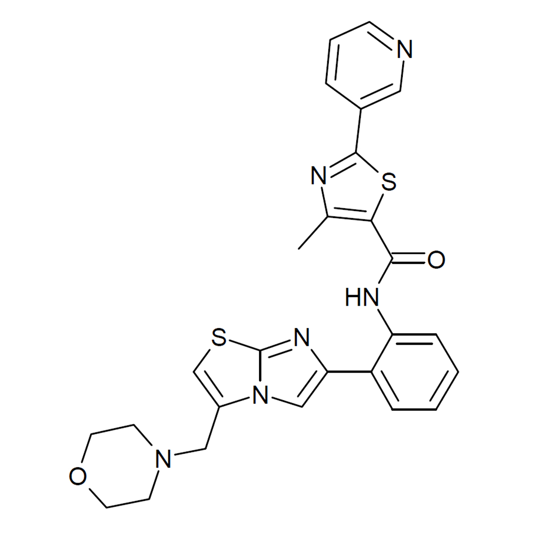
| IUPAC Name |
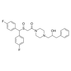
4-methyl-N-[2-[3-(morpholin-4-ylmethyl)imidazo[2,1-b][1,3]thiazol-6-yl]phenyl]-2-pyridin-3-yl-1,3-thiazole-5-carboxamide
|
| Molecular Formula | C₂₆H₂₄N₆O₂S₂ |
| Molecular Weight | 516.64 |
| Purity | ≥99% Pure (LC-MS) |
| Liquid Availability | N/A |
| Powder Availability |  1 gram |
| Gel Availability | N/A |
| Storage | Store in cool dry environment, away from direct sunlight. |
| Terms | All products are for laboratory developmental research USE ONLY. Products are not for human consumption. |
What is SRT2104?
SRT2104 is an experimental nootropic compound that has gained scientific interest due to its ability to activate sirtuin 1 (SIRT1), a protein involved in regulating cellular metabolism, inflammation, stress resistance, and aging-related processes. As a synthetic sirtuin-activating compound, SRT2104 was developed to mimic some of the biological effects associated with calorie restriction, a process linked to improved cellular function and longevity. Research suggests that SIRT1 activation may influence pathways important for brain health, including mitochondrial efficiency, oxidative stress reduction, neuronal survival, and cognitive resilience. While SRT2104 has been explored primarily for metabolic, inflammatory, and age-related conditions, its potential nootropic properties stem from its proposed ability to enhance cellular energy regulation and protect neurons from damage.
Main Research Findings
1) Administration of SRT2104 was found to elicit exercise mimetic effects that promoted recovery in cases of Duchenne muscular dystrophy.
2) Treatment with SRT2104 was shown to reduce diabetic-induced aortic endothelial dysfunction by inhibition of P53.
Selected Data
1) The study conducted by researchers Giovarelli et al investigated the therapeutic potential of SRT2104, a selective activator of silent information regulator 1 (SIRT1), for improving disease progression in Duchenne muscular dystrophy (DMD). The researchers designed a comprehensive experimental approach using multiple biological models, including dystrophic Drosophila melanogaster, mdx mice, and patient-derived human DMD muscle cells, to determine whether SRT2104 could restore muscle function, improve mitochondrial activity, reduce disease-related muscle degeneration, and reproduce beneficial effects typically associated with exercise. Because DMD is characterized by progressive skeletal muscle deterioration, mitochondrial dysfunction, inflammation, fibrosis, and impaired regeneration, the researchers focused on determining whether pharmacological activation of SIRT1 could influence these pathological mechanisms. SIRT1 was chosen as a target because it is a NAD+-dependent deacetylase involved in regulating mitochondrial metabolism, energy balance, inflammation, aging processes, and muscle adaptation. Compared with other SIRT1 activators such as resveratrol, SRT2104 was selected because it demonstrates greater potency, improved selectivity, better pharmacokinetic properties, and previous evidence of safety in clinical studies [1].
The first experimental model involved dystrophic Drosophila melanogaster carrying mutations in the dystrophin-like protein gene. The DysE17 fly model was used because dystrophin function is evolutionarily conserved and these animals develop progressive muscle impairment similar to aspects of human DMD. Wild-type Oregon-R flies served as controls. Adult flies between three and five days old were randomly assigned to treatment groups and maintained under controlled temperature and light conditions. SRT2104 was incorporated into the standard fly diet at concentrations ranging from 10 to 50 µg/mL, while control groups received vehicle-containing diets. Treatment continued for several weeks, and diets were replaced weekly. Researchers evaluated survival by recording the number of dead flies every five days and assessed muscle function using a climbing assay. In this test, flies were placed in empty vials, forced to climb after being tapped downward, and the percentage capable of reaching a designated height within sixty seconds was recorded. Muscle structure was examined using fluorescent phalloidin staining of flight muscles, while mitochondrial ultrastructure was analyzed through transmission electron microscopy [1].
To confirm the effects of SRT2104 in a mammalian system, researchers used mdx mice, a commonly used genetic model of DMD caused by dystrophin deficiency. The investigators generated dystrophic mice by crossing mdx animals with PhAM mice, allowing enhanced analysis of mitochondrial physiology. Age-matched control mice with identical genetic backgrounds were included. Male mdx mice at eight weeks of age received SRT2104 supplementation in their diet for twelve weeks. The compound was administered at a concentration of 1.33 g/kg of chow, resulting in an estimated daily dose of approximately 100 mg/kg body weight. Control mdx mice received the same diet without SRT2104. Animals were housed under standardized environmental conditions with unrestricted access to food and water. Body weight and food consumption were monitored throughout the study to evaluate possible toxicity.
Functional testing was performed to determine whether SRT2104 improved muscle strength and endurance. Whole-body tension testing measured the maximum force generated by mice during forward-pulling movements after tail stimulation. Multiple trials were recorded, and the strongest performances were averaged and normalized to body weight. Muscle endurance was assessed using a treadmill exhaustion test, where mice were trained to run before undergoing progressively increasing speeds until exhaustion occurred. Researchers recorded running time and distance to evaluate exercise capacity. Following functional assessments, muscle tissues including the diaphragm, tibialis anterior, and gastrocnemius were collected for additional analyses.
Histological and molecular techniques were used to examine the mechanisms responsible for SRT2104’s effects. Hematoxylin and eosin staining assessed general muscle morphology, while staining measured fibrosis by identifying collagen accumulation. Immunofluorescence techniques evaluated muscle membrane integrity, inflammation, regeneration, and maturation using markers such as CD45, embryonic myosin heavy chain, and MYOZ1. Muscle stem cells isolated from mdx mice were cultured and treated with SRT2104 to determine whether the compound directly improved myogenic differentiation. Human experiments were also performed using immortalized myoblast lines derived from pediatric DMD patients. These cells were treated with 3 μM SRT2104 during differentiation, and researchers evaluated myotube formation and mitochondrial membrane potential [1].
Finally, researchers investigated the molecular mechanisms underlying SRT2104’s effects through SIRT1 activity assays, molecular simulations, proteomics, acetylome profiling, and mitochondrial analyses. SIRT1 enzymatic activity was measured from muscle extracts, while computational modeling evaluated how SRT2104 altered SIRT1 structure and activation. Mass spectrometry approaches identified changes in protein abundance and lysine acetylation patterns associated with metabolism, muscle contraction, and energy production. Mitochondrial function was evaluated through measurements of ATP production, respiratory chain activity, oxygen consumption, and mitochondrial membrane potential. Statistical analyses were performed using blinded evaluation methods, with appropriate tests including t-tests and ANOVA depending on experimental comparisons. Overall, the methods provided a detailed evaluation of SRT2104 across multiple DMD models, allowing researchers to investigate functional, cellular, metabolic, and molecular outcomes related to SIRT1 activation [1].
2) The study completed by Wu et al investigated the ability of SRT2104, a selective small-molecule activator of sirtuin 1 (SIRT1), to protect against diabetes-induced aortic endothelial dysfunction and explored whether this protective mechanism occurs through regulation of P53 acetylation. Because endothelial dysfunction is a major contributor to diabetic macrovascular complications and is strongly associated with increased oxidative stress and inflammation, the researchers designed both in vivo and in vitro experiments to examine how activation of SIRT1 influences vascular injury caused by diabetes or high-glucose exposure. The study specifically focused on the relationship between SIRT1 activation and P53 because P53 is an important substrate of SIRT1, but the extent to which P53 deacetylation contributes to the vascular benefits of SIRT1 activation was previously unclear. SRT2104 was selected because it is a highly selective SIRT1 activator with greater specificity than earlier compounds such as resveratrol and has demonstrated safety in previous animal and human investigations [2].
For the animal experiments, researchers used eight-week-old male C57BL/6 mice housed under controlled environmental conditions with a 12-hour light/dark cycle and unrestricted access to food and water. Diabetes was induced through intraperitoneal injections of streptozotocin (STZ), a compound commonly used to create experimental diabetes by damaging pancreatic insulin-producing cells. Mice received STZ at 50 mg/kg/day for five consecutive days, while control animals received sodium citrate vehicle injections. One week after the final injection, fasting blood glucose levels were measured following a four-hour fast, and mice with glucose concentrations greater than 13.89 mM were classified as diabetic. After diabetes confirmation, animals were divided into groups receiving either a standard diet or a diet supplemented with SRT2104. SRT2104 was incorporated into chow at 1.33 g/kg, which was designed to deliver approximately 100 mg/kg/day. Blood glucose levels were monitored throughout the study at several time points to determine whether SRT2104 affected diabetic status [2].
To further evaluate whether P53 activation directly contributes to vascular dysfunction, an additional experiment was performed using healthy non-diabetic mice. These animals were treated with nutlin3a, a specific inhibitor of mouse double minute 2 (MDM2), which prevents normal P53 degradation and results in increased P53 activation. Eight-week-old male mice received intraperitoneal injections of nutlin3a at 10 mg/kg every other day for four weeks, while control animals received vehicle treatment. After completion of the experimental procedures, mice were anesthetized, sacrificed, and their aortas were collected for functional, molecular, and histological evaluation.
Aortic function was assessed by measuring vascular contractility because increased contraction is an important feature of diabetic endothelial dysfunction. Thoracic aortas were isolated, cut into 4 mm segments, and mounted onto metal supports connected to a force-displacement recording system. After equilibration, the tissues were exposed to increasing concentrations of phenylephrine (PE), a vasoconstrictor, ranging from 10⁻⁹ to 10⁻⁴ M. The force generated by the vessels was recorded to determine whether diabetes increased contractility and whether SRT2104 could restore normal vascular responses [2].
Histological and molecular analyses were performed to identify structural damage, oxidative stress, inflammation, SIRT1 expression, and P53 activation. Aortic tissues were fixed, embedded in paraffin, sectioned, and stained using hematoxylin and eosin (H&E) to evaluate changes in vascular morphology, including endothelial organization and tunica media thickness. Immunohistochemical staining was performed using markers of oxidative damage, including 3-nitrotyrosine (3-NT) and 4-hydroxynonenal (4-HNE), as well as inflammatory markers including vascular cell adhesion molecule-1 (VCAM-1) and intercellular adhesion molecule-1 (ICAM-1). Additional staining measured SIRT1 expression and acetylated P53 levels to evaluate the proposed pathway connecting SRT2104 treatment with P53 regulation.
Cell culture experiments were performed using endothelial cells isolated from mouse aortas. Cells were grown under normal glucose or high-glucose conditions to simulate the diabetic environment. To determine whether SRT2104 directly protected endothelial cells, high-glucose-treated cells were exposed to 3 μM SRT2104 for 48 hours. To determine whether P53 was necessary for SRT2104’s effects, researchers manipulated P53 activity using two approaches. First, P53 expression was reduced using P53-specific siRNA, allowing researchers to compare the effects of P53 inhibition with SRT2104 treatment. Second, P53 activity was increased using nutlin3a to determine whether forced P53 activation could prevent the protective effects of SRT2104 [2].
Additional molecular experiments included real-time PCR, Western blotting, and biochemical assays. Real-time PCR measured inflammatory gene expression, including Vcam-1, Icam-1, and Mcp-1. Western blot analysis quantified proteins such as SIRT1, total P53, acetylated P53, and inflammatory markers. Oxidative stress was evaluated by measuring reactive oxygen species (ROS) and malondialdehyde (MDA), a marker of lipid oxidation. Data were analyzed statistically using Student’s t-tests and one-way ANOVA. Overall, the experimental design allowed the researchers to evaluate the vascular protective effects of SRT2104 while determining whether inhibition of P53 activity was required for its mechanism of action [2].
Discussion
1) The results of the study by Giovarelli et al demonstrated that SRT2104 produced significant therapeutic benefits across multiple models of Duchenne muscular dystrophy by improving muscle performance, enhancing mitochondrial metabolism, reducing pathological muscle changes, and creating molecular adaptations similar to those produced by exercise. Initial experiments using dystrophic Drosophila melanogaster showed that SRT2104 improved muscle function without causing harmful effects. Survival analysis demonstrated that SRT2104 treatment did not negatively affect the lifespan of dystrophic flies, indicating that the compound was well tolerated. Although untreated DysE17 flies developed progressive movement impairment, SRT2104-treated flies showed preservation of climbing ability over time. The decline in mobility occurred more slowly in treated animals, suggesting that SRT2104 reduced the progression of dystrophic muscle dysfunction. Examination of flight muscle structure revealed that untreated dystrophic flies displayed disrupted muscle organization, abnormal spaces between fibers, and increased areas of tissue degeneration. Treatment with SRT2104 partially restored normal muscle architecture and significantly reduced damaged muscle regions. Transmission electron microscopy further showed that untreated dystrophic flies had abnormal mitochondria with damaged structures and disrupted cristae, while SRT2104 improved mitochondrial organization [1].
The beneficial effects observed in flies were confirmed in the mdx mouse model of DMD. Twelve weeks of dietary SRT2104 treatment produced no evidence of toxicity, as treated mice showed normal body weight progression and food intake compared with untreated controls. Functional assessments revealed that SRT2104 significantly enhanced muscle performance. During treadmill testing, SRT2104-treated mdx mice ran longer distances and demonstrated increased time before exhaustion compared with untreated mdx mice, indicating improved exercise tolerance. Whole-body tension testing also demonstrated increased muscle force production after treatment. Importantly, improvements in strength appeared before the end of the treatment period, with measurable benefits observed after six weeks and maintained throughout the twelve-week intervention.
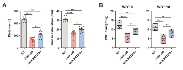
Figure 1: Effects of treatment with SRT2104 on A) distance ran, and B) whole body tension in varying strains of mice.
Histological analysis showed that SRT2104 reduced several major features of dystrophic muscle damage. The diaphragm, which is severely affected in DMD and contributes to respiratory failure, showed improved tissue organization after treatment. Compared with untreated mdx mice, SRT2104-treated animals demonstrated fewer necrotic muscle fibers and reduced immune cell infiltration, shown by decreased CD45-positive inflammatory cells. Similar protective effects were also observed in tibialis anterior muscle, indicating that the benefits were not limited to one muscle group. Fibrosis analysis showed that SRT2104 reduced collagen accumulation and lowered expression of fibrotic and adipogenic genes, suggesting that the compound slowed replacement of functional muscle with scar-like tissue [1].
SRT2104 also enhanced muscle regeneration. Treated mdx mice demonstrated increased numbers of regenerating fibers positive for embryonic myosin heavy chain, indicating activation of repair processes. In addition, expression of MYOZ1, a marker associated with mature muscle fibers, increased after treatment, suggesting that SRT2104 not only promoted early regeneration but also supported the development of functional muscle fibers. Muscle stem cell experiments confirmed these findings by showing that SRT2104 improved the ability of dystrophic muscle cells to fuse and form mature myotubes. These results indicated that SRT2104 preserved the regenerative potential of muscle cells under dystrophic conditions [1].
Mechanistic experiments confirmed that SRT2104 enhanced SIRT1 activity without changing overall SIRT1 protein levels. Molecular simulations suggested that SRT2104 promoted a structural shift of SIRT1 into its active configuration, allowing increased enzymatic function. Increased SIRT1 activation resulted in widespread changes in protein acetylation patterns. Acetylome analysis revealed that SRT2104 altered proteins involved in skeletal muscle contraction, ATP production, glycolysis, and mitochondrial activity. Many of these molecular changes resembled adaptations normally produced by repeated exercise, supporting the conclusion that SRT2104 acts as an exercise mimetic compound.
Proteomic and metabolic analyses further showed that SRT2104 improved energy metabolism in dystrophic muscle. Treatment increased proteins involved in fatty acid oxidation while modifying pathways associated with glucose metabolism, suggesting a shift toward more efficient energy utilization. SRT2104-treated muscles demonstrated higher ATP levels and improved mitochondrial respiratory capacity, particularly through enhanced complex II-driven respiration. These improvements occurred without increasing mitochondrial abundance, indicating that SRT2104 primarily enhanced mitochondrial quality and function rather than simply increasing mitochondrial quantity [1].
Experiments using human DMD patient-derived myoblasts supported the therapeutic relevance of the findings. Human dystrophic muscle cells normally displayed impaired differentiation and reduced mitochondrial function; however, SRT2104 treatment improved myotube formation and restored mitochondrial membrane potential. Overall, the study demonstrated that SRT2104 counteracted several central mechanisms of DMD progression, including mitochondrial dysfunction, inflammation, fibrosis, impaired regeneration, and reduced muscle performance. The researchers concluded that SRT2104 represents a promising therapeutic candidate capable of producing exercise-like adaptations in dystrophic muscle through activation of SIRT1-dependent pathways [1].
2) The results of the study completed by Wu et al demonstrated that SRT2104 significantly protected against diabetes-induced aortic endothelial dysfunction by activating SIRT1 and reducing P53-mediated oxidative stress and inflammation. Initial findings confirmed that streptozotocin successfully produced diabetes in mice, as diabetic animals developed significantly elevated blood glucose levels compared with control animals. Treatment with SRT2104 did not significantly change blood glucose concentrations in either diabetic or non-diabetic mice, indicating that its vascular benefits were not caused by improvements in glucose control. Instead, SRT2104 appeared to directly influence vascular signaling pathways responsible for endothelial protection. When aortic contractility was evaluated using phenylephrine stimulation, diabetic mice demonstrated significantly increased vascular contraction compared with healthy controls, confirming the presence of endothelial dysfunction. However, diabetic mice treated with SRT2104 showed a major reduction in this exaggerated contractile response, demonstrating that SRT2104 improved vascular function despite persistent hyperglycemia [2].
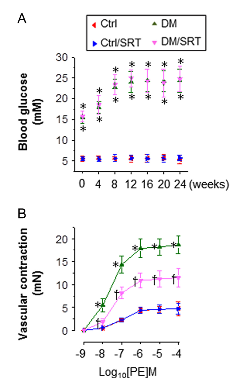
Figure 2: Changes in A) blood glucose levels, and B) vascular contraction following treatment with SRT2104.
Structural analysis of aortic tissue further supported the protective effects of SRT2104. Hematoxylin and eosin staining revealed that diabetic mice experienced mild thickening of the tunica media layer and disruption of endothelial and smooth muscle cell organization. Although diabetes did not cause severe structural abnormalities, these changes represented early vascular remodeling associated with diabetic complications. SRT2104 treatment reduced tunica media thickening and improved vascular structure, suggesting that activation of SIRT1 helped prevent diabetes-related vascular remodeling.
The researchers next examined whether SRT2104 reduced oxidative stress and inflammation, two major mechanisms contributing to diabetic vascular damage. Diabetic mice demonstrated increased levels of oxidative stress markers, including 3-nitrotyrosine and 4-hydroxynonenal, within the aortic wall. These markers indicated increased protein nitration and lipid oxidation caused by excessive reactive oxygen species production. SRT2104 treatment significantly decreased both markers, demonstrating reduced oxidative damage. Inflammatory signaling was also increased in diabetic aortas, as shown by elevated VCAM-1 and ICAM-1 expression. These adhesion molecules promote immune cell attachment and contribute to vascular inflammation. Treatment with SRT2104 significantly reduced both VCAM-1 and ICAM-1 protein expression, while also decreasing inflammatory gene transcription, confirming its anti-inflammatory effects [2].
Evaluation of the SIRT1/P53 pathway revealed that diabetes reduced vascular SIRT1 expression while causing increased P53 acetylation, a modification associated with greater P53 stability and activity. SRT2104 reversed these diabetes-induced changes by increasing SIRT1 levels and greatly decreasing acetylated P53 expression. In diabetic mice, SRT2104 increased SIRT1 protein approximately 3.79-fold and reduced acetylated P53 by more than 80%, demonstrating strong activation of the SIRT1-dependent P53 regulatory pathway. These findings suggested that SRT2104 protects endothelial cells by restoring SIRT1 activity and preventing excessive activation of P53 [2].
Cell culture experiments confirmed these findings at the cellular level. Endothelial cells exposed to high glucose showed reduced SIRT1 expression, increased acetylated P53, elevated ROS and MDA production, and increased expression of inflammatory genes such as Vcam-1 and Mcp-1. Treatment with SRT2104 reversed these effects by restoring SIRT1 expression, reducing P53 acetylation, decreasing oxidative stress, and suppressing inflammatory signaling. These findings demonstrated that SRT2104 directly acts on endothelial cells rather than producing benefits only through systemic changes in animals.
Further experiments demonstrated that P53 was a critical mediator of SRT2104’s protective effects. When P53 expression was reduced using P53 siRNA, endothelial cells showed similar improvements to those produced by SRT2104, including increased SIRT1 expression and reduced oxidative stress and inflammation. Importantly, adding SRT2104 after P53 silencing did not provide additional protection, suggesting that P53 inhibition was the primary pathway responsible for SRT2104’s effects. Conversely, when P53 was activated using nutlin3a, the protective effects of SRT2104 were eliminated. High-glucose-treated endothelial cells exposed to both SRT2104 and nutlin3a continued to demonstrate oxidative stress and inflammatory activation, confirming that P53 deacetylation was required for SRT2104-mediated protection [2].
Finally, experiments using healthy mice showed that forced activation of P53 alone was sufficient to produce vascular dysfunction. Nutlin3a treatment increased acetylated P53 levels, promoted oxidative damage, increased inflammatory marker expression, and enhanced phenylephrine-induced aortic contraction even without diabetes. These findings established P53 as an important contributor to endothelial dysfunction rather than simply a secondary response to diabetes. Overall, the study concluded that SRT2104 protects against diabetes-induced vascular injury primarily by activating SIRT1, which reduces P53 acetylation and prevents oxidative stress, inflammation, and abnormal vascular contraction. The findings identify the SIRT1-P53 pathway as a promising therapeutic target for preventing diabetic vascular complications [2].
Disclaimer
**LAB USE ONLY**
*This information is for educational purposes only and does not constitute medical advice. THE PRODUCTS DESCRIBED HEREIN ARE FOR RESEARCH USE ONLY. All clinical research must be conducted with oversight from the appropriate Institutional Review Board (IRB). All preclinical research must be conducted with oversight from the appropriate Institutional Animal Care and Use Committee (IACUC) following the guidelines of the Animal Welfare Act (AWA).
Citations
[1] Giovarelli M, Zecchini S, Casati SR, et al. The SIRT1 activator SRT2104 exerts exercise mimetic effects and promotes Duchenne muscular dystrophy recovery. Cell Death Dis. 2025;16(1):259. Published 2025 Apr 7. doi:10.1038/s41419-025-07595-z
[2] Wu H, Wu J, Zhou S, et al. SRT2104 attenuates diabetes-induced aortic endothelial dysfunction via inhibition of P53. J Endocrinol. 2018;237(1):1-14. doi:10.1530/JOE-17-0672
SRT2104 is sold for laboratory research use only. Terms of sale apply. Not for human consumption, nor medical, veterinary, or household uses. Please familiarize yourself with our Terms & Conditions prior to ordering.


| File Name | View/Download |
| Coming Soon! |  |
VIEW CERTIFICATES OF ANALYSIS (COA)

Reviews
There are no reviews yet.