


Description
Urolithin B Nootropic Powder (50 Grams)






| CAS Number | 1139-83-9 |
| Other Names | UrolithinB, Urolithin B, Urolithin-B |
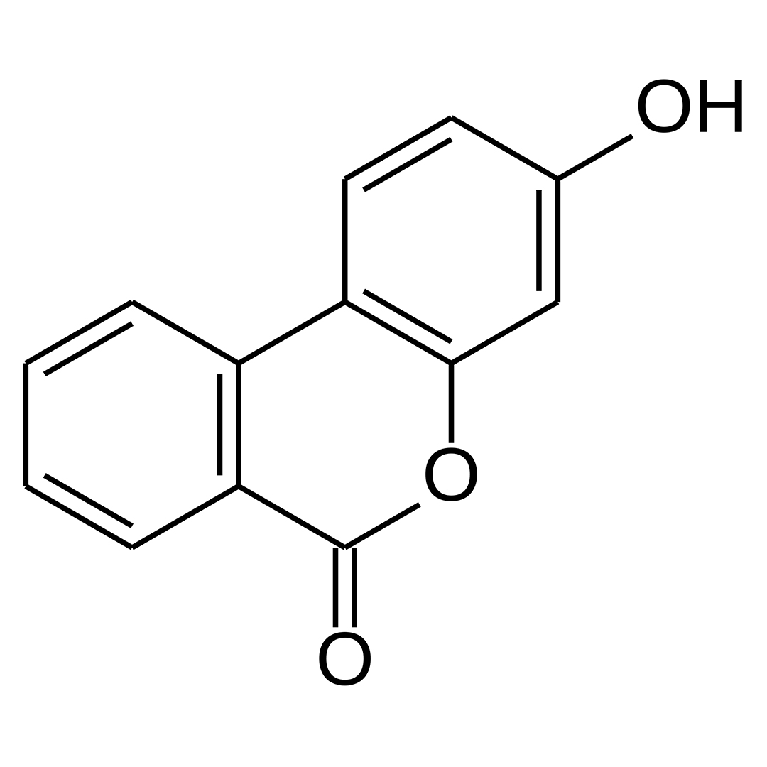
| IUPAC Name | 3-Hydroxy-6H-dibenzo[b,d]pyran-6-one |
| Molecular Formula | C₁₃H₈O₃ |
| Molecular Weight | 212.20 |
| Purity | ≥99% Pure (LC-MS) |
| Powder Availability |  60 capsules (150mg/capsule, 9,000mg total) |
| Storage | Store in cool dry environment, away from direct sunlight. |
| Terms | All products are for laboratory developmental research USE ONLY. Products are not for human consumption. |
What is Urolithin B?
Urolithin B is a gut-derived metabolite produced from dietary ellagitannins that has attracted interest for its potential neuroprotective and cognitive-supporting properties. Preclinical studies indicate it may enhance mitochondrial function, reduce neuroinflammation, and promote cellular mitophagy. These processes act as mechanisms that could support brain health, resilience, and cognitive performance.
Main Research Findings
1) Through the inhibition of the ERK/NF-kB pathways, Urolithin B was found to suppress the activation of osteoclasts and reduce osteoporosis-related bone loss.
2) By modulating Nrf2-regulated antioxidation, Urolithin B was able to attenuate cerebral ischemia-reperfusion injury in an animal model of research.
Selected Data
1) This comprehensive study completed by the research team of Li et al aimed to investigate the inhibitory effects of Urolithin B (UB) on osteoclastogenesis and bone loss, both in vitro and in vivo, particularly focusing on its impact on the ERK/NF-κB signaling pathway. The research commenced with an in vitro phase, utilizing two primary cell models: bone marrow-derived macrophages (BMMs) isolated from 6-week-old C57BL/6 mice and RAW264.7 cells, a murine macrophage cell line. All cells were maintained under standard culture conditions at 37°C with 5% CO2 in DMEM medium supplemented with 10% FBS and antibiotics [1].
Initial experiments focused on assessing the cytotoxicity of UB using the CCK-8 assay. BMMs and RAW264.7 cells were seeded in 96-well plates and exposed to various concentrations of UB, including 0, 1, 5, 25, 50, 100, and 150 µmol/L, for 1, 2, or 3 days. Cell viability was determined by measuring absorbance at 450 nm, with concentrations showing minimal toxicity selected for subsequent experiments. Osteoclast differentiation in vitro was induced by treating BMMs with 50 ng/mL M-CSF and 50 ng/mL RANKL, critical cytokines for osteoclast formation. During this differentiation process, cells were concurrently treated with different concentrations of UB including 0, 1, 5, and 25 µmol/L, for 5 to 7 days, depending on the specific assay.
To evaluate the extent of osteoclast formation and function, several detailed in vitro assays were employed. Tartrate-resistant acid phosphatase (TRAP) staining was performed on differentiated BMMs to quantify the number and average area of multi-nucleated osteoclasts, which are characteristic of mature osteoclasts. F-actin staining and immunofluorescence were used to visualize the morphology of osteoclasts, specifically the formation of actin rings essential for bone resorption, and to assess the expression of functional proteins such as MMP9. DAPI staining was used to identify cell nuclei. The bone resorption capabilities of osteoclasts were directly measured using a Pit Formation Assay, where BMMs were cultured on osteo-assay plates. After UB treatment, the bone resorption pits were imaged and quantitatively analyzed using IMAGEJ software to determine the resorbed area [1].
Molecular analyses included Western blot and quantitative RT-PCR to investigate the expression of osteoclast-related protein genes and key signaling molecules. Western blotting was performed on RAW264.7 cells treated with RANKL and various UB concentrations, allowing for the analysis of proteins like MMP9, CTSK, c-fos, and NFATc1. Furthermore, the study meticulously examined the RANKL-mediated activation of the MAPK/NF-κB pathway by Western blot, assessing the phosphorylation and degradation levels of IκB and P65, both NF-κB pathway components, as well as the phosphorylation levels of ERK, JNK, and p38, MAPK pathway components, at various time points including 0, 5, 15, 30, 60 min, following intervention. Specific pathway inhibitors, SCH772984 for ERK and SC75741 for P65, were also utilized to further dissect the involvement of these pathways. Quantitative RT-PCR was used to analyze the mRNA expression levels of osteoclast-related genes, MMP9, CTSK, TRAP, V-ATPase, c-fos, and NFATc1, in BMMs under similar treatment conditions.
For the in vivo component, a well-established ovariectomized (OVX) mouse model of osteoporosis was utilized. Forty 6-week-old C57BL/6 mice were randomly divided into four groups: sham-operated control, OVX vehicle control, OVX + low-dose UB of 10 mg/kg, and OVX + high-dose UB of 50 mg/kg. OVX surgery involved bilateral ovariectomy, while sham-operated mice underwent a similar procedure without ovary removal. Three weeks post-surgery, UB was administered via intraperitoneal injection every two days for 8 weeks. After the intervention period, blood samples were collected to measure serum C-telopeptide of type I collagen (CTX-1), a bone metabolism marker, using an ELISA kit. The femurs of the mice were harvested and fixed for morphological and histological analyses [1].
Micro-CT scanning was employed to obtain high-resolution 3D images of the left femurs. Detailed structural parameters of the bone were measured and analyzed using CTAn software, including bone mineral density (BMD), bone surface area (BS), bone volume (BV), bone volume to total volume ratio (BV/TV), bone surface to bone volume ratio (BS/BV), trabecular separation (Tb.Sp), and number of trabecular bone (Tb.N). Histological analyses involved decalcifying and embedding femurs in paraffin, followed by sectioning and staining with Hematoxylin and Eosin (H&E) to visualize overall bone morphology and TRAP staining to quantify osteoclast presence in bone tissues. Immunohistochemical staining was performed to detect and quantify MMP9 and NFATc1 positive cells in the bone sections, providing further insights into osteoclast activity in vivo. All experimental data were expressed as mean ± standard deviation, and statistical significance was determined using t-tests and one-way ANOVA [1].
2) This study completed by the research team of Li et al explored the neuroprotective effects of Urolithin B (UB) against cerebral ischemia-reperfusion injury (IRI) in rats, focusing on its modulation of the Nrf2-regulated anti-oxidation pathway. The study primarily employed in vivo animal models, specifically male Sprague-Dawley rats weighing 180–220 g. All animals were housed under controlled environmental conditions, including a consistent temperature, humidity, and a 12-hour light/dark cycle, with ad libitum access to food and water, followed by a 7-day acclimatization period [2].
The core experimental model involved inducing middle cerebral artery occlusion (MCAO) surgically in the rats, a well-established method for cerebral ischemia. This procedure entailed isolating and ligating the common carotid artery (CCA) and the right external carotid artery (ECA). A monofilament nylon thread, featuring a rounded silica gel tip with a diameter of 0.28 ± 0.02 mm, was then carefully inserted through a small incision in the CCA, extending approximately 18-20 mm into the right anterior cerebral artery. After a 2-hour period of ischemia, the nylon thread was gently withdrawn to initiate reperfusion. Sham-operated animals underwent the same procedure, but without thread insertion. Body temperature was consistently maintained at 37 °C throughout the surgery using a heating pad. The rats were sacrificed 24 hours after reperfusion [2].
Animals were randomly assigned to one of four groups, each comprising five rats: a Sham group receiving vehicle injection, an IR group receiving vehicle injection, an IR + UB group, and an IR + UB + ATRA group. UB was administered via intraperitoneal injection at a concentration of 0.07 mg/kg, dissolved in a mixture of 2% DMSO and normal saline, one hour before MCAO surgery. All-trans retinoic acid (ATRA), an Nrf2 inhibitor, was administered at 5 mg/kg, also via intraperitoneal injection, alongside UB in the IR + UB + ATRA group, one hour before MCAO. Vehicle injections of 2% DMSO in normal saline were volume-matched across all groups to ensure consistency.
To assess the impact of these treatments, a battery of functional and histological evaluations was conducted. Neurological deficits were evaluated before sacrificing the rats using the modified Longa standard, classifying impairment from Grade 0 (no impairment) to Grade 4 (complete loss of autonomous walking). Cerebral infarct volume was measured using 2,3,5-triphenyltetrazolium chloride (TTC) staining. After cardiac perfusion with normal saline and careful brain extraction, brains were frozen, sectioned into 2 mm slices, and incubated in 2% TTC solution at 37 °C. Post-fixation with 4% paraformaldehyde, images were captured and analyzed using ImageJ software, applying Swanson’s method to account for edema.
Neuronal damage and apoptosis were assessed through Nissl staining, TUNEL assay, and immunohistochemistry for Caspase-3. For Nissl staining, paraffin-embedded brain sections were stained with Cresyl violet to count surviving neurons with intact morphology in the ischemic hemisphere’s parietal cortex and hippocampal CA1 sector. TUNEL staining was performed on paraffin-embedded sections after proteinase K treatment to identify apoptotic cells, followed by hematoxylin counterstaining. Immunohistochemistry for Caspase-3, a marker of apoptosis, was carried out using a primary anti-caspase-3 antibody and an enhanced secondary antibody, with positive cells quantified [2].
Oxidative stress levels were comprehensively analyzed. Biochemical assays were performed on dissected and homogenized ischemic ipsilateral parietal cortex and hippocampal CA1 sector. Malondialdehyde (MDA) concentration, an indicator of lipid peroxidation, and superoxide dismutase (SOD) activity, a measure of antioxidant defense, were quantified using specific detection kits. Immunohistochemistry was also used to detect 8-hydroxyl-2′-deoxyguanosine (8-OHdG), a marker of oxidative DNA damage, and inducible nitric oxide synthase (iNOS), involved in reactive nitrogen species production, using anti-8-OHdG and anti-iNOS primary antibodies, respectively.
The study further investigated the Nrf2/HO-1 signaling pathway using immunofluorescence, qRT-PCR, and Western blot. Immunofluorescence staining was performed on brain sections using anti-Nrf2 and anti-HO-1 antibodies, followed by CY3-conjugated secondary antibodies and Hoechst stain, to visualize the nuclear localization of these proteins. Total RNA was extracted from the cerebral cortex for qRT-PCR to measure mRNA levels of Nrf2 and HO-1 using specific primers. Western blot analysis was conducted on protein extracts from the ischemic ipsilateral parietal cortex and hippocampus using anti-Nrf2 and anti-HO-1 primary antibodies to quantify protein expression levels. Statistical analyses were performed using one-way ANOVA followed by Bonferroni’s test [2].
Discussion
1) The results of the study by Li et al investigating Urolithin B (UB)’s impact on osteoclast activation and bone loss yielded significant findings across both in vitro and in vivo experiments. Initially, cell viability assays demonstrated that UB exhibited minimal cytotoxicity at concentrations up to 25 µmol/L in both BMMs and RAW264.7 cells, making these concentrations suitable for subsequent in vitro studies on osteoclast differentiation and function. However, concentrations exceeding 50 µmol/L significantly affected cell viability, guiding the selection of experimental doses [1].
In the in vitro assessment of osteoclast differentiation, UB effectively inhibited RANKL-induced osteoclast formation in a concentration-dependent manner. TRAP staining, a hallmark assay for osteoclasts, revealed a marked reduction in both the number and average area of osteoclasts in BMMs treated with UB compared to the RANKL-induced control group. This inhibitory effect became increasingly pronounced with higher UB concentrations of 1, 5, and 25 µmol/L. Further visualization using immunofluorescence staining corroborated these findings, showing that UB treatment significantly suppressed the formation of F-actin rings, critical structures for osteoclast adhesion and bone resorption. Moreover, the number of nuclei per osteoclast was significantly reduced in the presence of UB, indicating impaired osteoclast maturation. The expression of functional osteoclast-related proteins, such as MMP9, was also observed to be clearly inhibited by UB treatment in vitro [1].
The study then directly assessed UB’s effect on osteoclast bone resorption function using the Pit Formation Assay. The results unequivocally demonstrated that UB significantly suppressed the formation of bone resorption pits. Quantitatively, the resorbed hydroxyapatite area was reduced in a dose-dependent fashion, with UB doses of 1, 5, and 25 µmol/L decreasing the resorbed area from 51.25% in the RANKL group to 29.44%, 18.83%, and 4.84%, respectively, highlighting its potent anti-resorptive properties.
At the molecular level, Western blot analysis revealed that UB downregulated the expression of key osteoclast-related proteins, including MMP9, CTSK, c-fos, and NFATc1, in a concentration-dependent manner in RAW264.7 cells. Time-course experiments further indicated that UB reversed the RANKL-induced increase in these proteins, showing significant inhibitory effects after 12 hours, 1 day, and 3 days of intervention. Quantitative RT-PCR experiments confirmed these protein-level observations, demonstrating a significant decrease in the mRNA expression levels of osteoclast-related genes MMP9, CTSK, TRAP, V-ATPase, c-fos, and NFATc1, in BMMs treated with UB, reinforcing its role in inhibiting osteoclast gene transcription [1].
A crucial aspect of the study was elucidating the underlying signaling pathways. Western blot analysis showed that UB repressed RANKL-induced osteoclastogenesis by modulating both the NF-κB and MAPK pathways. Specifically, UB was found to significantly decrease the RANKL-induced phosphorylation and subsequent degradation of IκBα, an inhibitory protein of NF-κB, and the phosphorylation of P65, a key component of the NF-κB pathway. The peak phosphorylation of P65, which occurred rapidly after RANKL addition, was notably attenuated and delayed in UB-treated cells. Concurrently, UB significantly down-regulated the phosphorylation levels of ERK, a critical component of the MAPK pathway, while also showing inhibitory trends for p-JNK and p-p38. These inhibitory effects on ERK and P65 phosphorylation were observed to be concentration-dependent. To validate these pathway involvements, specific inhibitors were used: SCH772984, an ERK inhibitor, significantly inhibited ERK phosphorylation and synergistically enhanced UB’s reduction of CTSK and NFATc1 expression. Similarly, SC75741, a P65 inhibitor, decreased P65 phosphorylation and osteoclast-related protein expression, although UB alone demonstrated a more potent therapeutic effect than SC75741. When combined, UB and SC75741 showed an even greater reduction in osteoclast-related proteins [1].
The in vivo experiments confirmed UB’s therapeutic potential in an OVX-induced osteoporosis mouse model. Micro-CT analysis of the femurs revealed that UB treatment significantly improved several bone structural parameters that were compromised in OVX mice. Specifically, UB increased BMD, BV, and the ratio of bone volume to total volume (BV/TV), while decreasing Tb.Sp and increasing the number of Tb.N in a concentration-dependent manner. Histological examination with H&E staining further demonstrated that UB treatment decreased OVX-induced bone mass loss. TRAP staining of bone tissue sections showed a significant reduction in the number of TRAP-positive osteoclasts per bone surface in UB-treated groups, confirming its inhibitory effect on osteoclast activity in vivo.
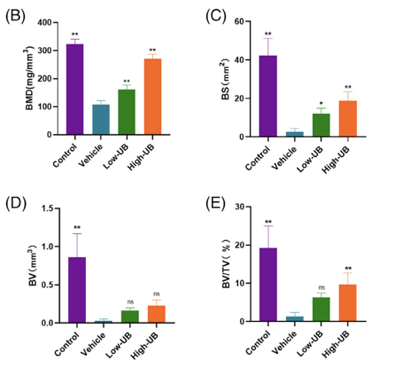
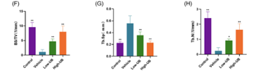
Figure 1: Changes in B) BMD, C) BS, D) BV, E) BV/TV, F) BS/TV, G) Tb.Sp, and H) Tb.N in response to treatment with a vehicle, a low dose of UB, or a high dose of UB.
Immunohistochemical analysis further supported these findings, revealing fewer MMP9 and NFATc1 positive cells in the bone tissue of UB-treated mice compared to the OVX control. Finally, serum analysis showed that UB treatment significantly reduced the content of CTX-1, a key bone resorption marker, in the serum of OVX mice. Importantly, no significant signs of drug toxicity were observed in the liver and kidney sections of UB-treated mice, suggesting a favorable safety profile. Collectively, these results strongly indicate that UB attenuates bone loss in OVX mice by inhibiting osteoclast formation and activity via the downregulation of the ERK/NF-κB signaling pathway [1].
2) The results of this study conducted by Li et al demonstrated that UB treatment significantly attenuates cerebral IRI in rats by modulating the Nrf2-regulated anti-oxidation pathway. The initial assessment of infarct volume using TTC staining revealed a dramatic protective effect of UB. While the Sham and Sham + UB groups showed no tissue infarction, the IR + vehicle group exhibited extensive infarct areas, with an average infarct ratio of 48.14% ± 8.22%. Remarkably, UB administration before MCAO significantly reduced this infarct volume to a mere 4.34% ± 2.32%, highlighting its potent ability to limit tissue damage [2].
Consistent with the reduction in infarct volume, UB treatment also led to a significant improvement in neurological function. Rats in the IR + vehicle group displayed severe neurological deficits, indicated by a score of 1.80 ± 0.45. In contrast, the IR + UB group showed a significantly improved neurological deficit score of 0.40 ± 0.54, approaching the normal function observed in the sham group. These findings collectively underscored UB’s capacity to mitigate IR-induced functional impairments.
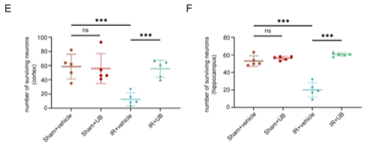
Figure 2: Number of surviving neurons within E) the cortex and F) the hippocampus following treatment with the experimental compound.
Further microscopic examination corroborated these neuroprotective effects. Nissl staining revealed severe neuronal damage in the IR + vehicle group, characterized by a significant reduction in Nissl bodies and extensive morphological shrinkage of neurons, with surviving neuron counts of 12.6 ± 9.29 in the cortex and 20.00 ± 8.43 in the hippocampus. UB treatment, however, substantially preserved neuronal integrity, increasing surviving neuron counts to 55.60 ± 12.28 in the cortex and 60.60 ± 1.82 in the hippocampus, effectively preventing widespread neuronal loss [2].
UB’s ability to inhibit neuronal apoptosis was a key finding. TUNEL staining showed a high percentage of apoptotic cells in the IR + vehicle group, measured at 35.0% ± 3.5% in the cortex and 35.7% ± 1.4% in the hippocampus. UB treatment significantly reduced these apoptotic cell percentages to 13.4% ± 1.5% in the cortex and 11.4% ± 0.9% in the hippocampus. Similarly, immunohistochemistry for Caspase-3, a crucial apoptotic effector, confirmed these results. The IR + vehicle group displayed high Caspase-3 positive ratios measured at 32.1% ± 2.0% in the cortex and 30.6% ± 1.3% in the hippocampus, which were markedly reduced by UB administration to 14.0% ± 1.0% in the cortex and 11.4% ± 0.9% in the hippocampus. These results firmly established UB as an effective inhibitor of neuronal apoptosis following IRI [2].
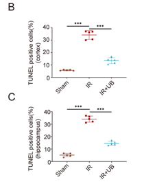
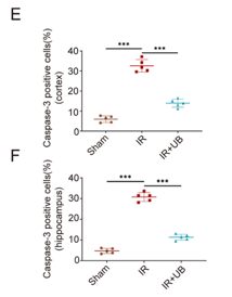
Figure 3: Changes in the number of TUNEL positive cells in B) the cortex and C) the hippocampus, as well as changes in the number of Caspase-3 positive cells in E) the cortex and F) the hippocampus.
A crucial aspect of UB’s neuroprotective mechanism was its significant alleviation of oxidative stress. Immunohistochemical analysis demonstrated that UB treatment substantially decreased the number of 8-OHdG-positive cells, a marker of oxidative DNA damage, and concurrently downregulated iNOS levels, which are associated with reactive nitrogen species production. Biochemical assays further supported these findings: UB significantly reduced the elevated levels of MDA, a marker of lipid peroxidation, in both the cortex and hippocampus of IR rats. Concomitantly, UB treatment restored and significantly increased SOD activity, a critical endogenous antioxidant enzyme, indicating an enhancement of the brain’s antioxidant defense system.
The study further elucidated the molecular mechanism underlying UB’s antioxidant effects, identifying the Nrf2/HO-1 signaling pathway as central. Immunofluorescence analysis clearly showed that UB treatment promoted the nuclear localization of both Nrf2 and HO-1 in neurons, indicating activation of this pathway. Quantitative RT-PCR confirmed a significant increase in mRNA expression levels of both Nrf2 by 3.40 ± 0.05 vs 2.15 ± 0.14 in IR+vehicle, and HO-1 by 8.54 ± 0.15 vs 4.11 ± 0.34 in IR+vehicle in the cerebral cortex of UB-treated rats. Western blot analysis consistently mirrored these findings, showing higher protein expression levels of Nrf2 by 2.34 ± 0.18 vs 1.84 ± 0.20, and HO-1 by 3.81 ± 0.23 vs 2.08 ± 0.22 in UB-treated groups [2].
The critical role of the Nrf2 pathway was definitively confirmed through experiments using ATRA, a specific Nrf2 inhibitor. When ATRA was co-administered with UB, the neuroprotective effects of UB were largely abrogated. Rats in the IR + UB + ATRA group exhibited a significant increase in neurological deficit scores and infarct ratios compared to the IR + UB group. Moreover, ATRA pretreatment substantially inhibited the anti-apoptotic effect of UB, as evidenced by a notable increase in TUNEL-positive cells. Molecular analyses in the ATRA group further revealed a significant decrease in Nrf2 and HO-1 expression at both mRNA and protein levels, and a notable reduction in their nuclear localization. These findings collectively provided compelling evidence that UB exerts its neuroprotective effects against IRI primarily by activating the Nrf2/HO-1 signaling pathway, thereby boosting antioxidant defenses and reducing oxidative damage and apoptosis [2].
Disclaimer
**LAB USE ONLY**
*This information is for educational purposes only and does not constitute medical advice. THE PRODUCTS DESCRIBED HEREIN ARE FOR RESEARCH USE ONLY. All clinical research must be conducted with oversight from the appropriate Institutional Review Board (IRB). All preclinical research must be conducted with oversight from the appropriate Institutional Animal Care and Use Committee (IACUC) following the guidelines of the Animal Welfare Act (AWA).
Citation
[1] Li Y, Zhuang Q, Tao L, et al. Urolithin B suppressed osteoclast activation and reduced bone loss of osteoporosis via inhibiting ERK/NF-κB pathway. Cell Prolif. 2022;55(10):e13291. doi:10.1111/cpr.13291
[2] Li ZW, Tang H, Chen XX, et al. Urolithin B Attenuates Cerebral Ischemia-reperfusion Injury by Modulating Nrf2-regulated Anti-oxidation in Rats. Neuroscience. 2024;538:46-58. doi:10.1016/j.neuroscience.2023.11.002
Urolithin B is sold for laboratory research use only. Terms of sale apply. Not for human consumption, nor medical, veterinary, or household uses. Please familiarize yourself with our Terms & Conditions prior to ordering.


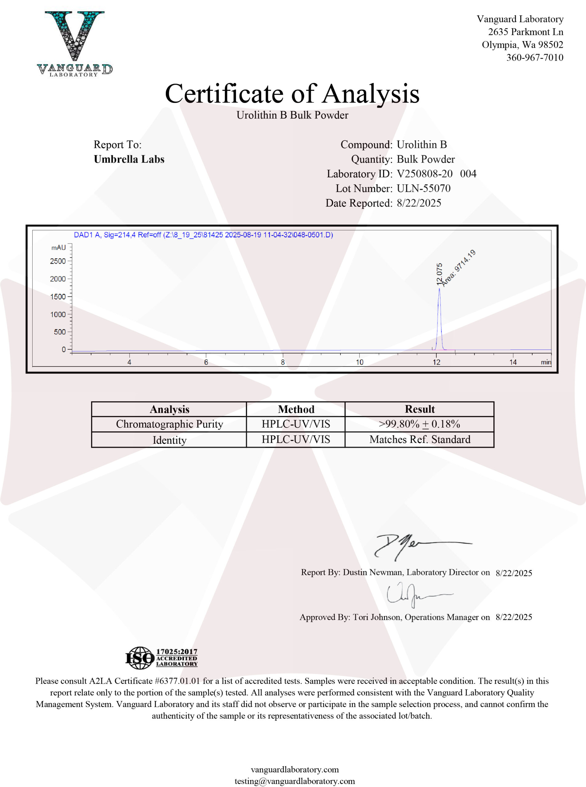
| File Name | View/Download |
| 2025-08-22-Umbrella-Labs-Urolithin-B-Certificate-of-Analysis-COA.pdf |  |
VIEW CERTIFICATES OF AUTHENTICITY (COA)


Reviews
There are no reviews yet.