
Description
Vitamin B3 Nootropic Powder (50 Grams)







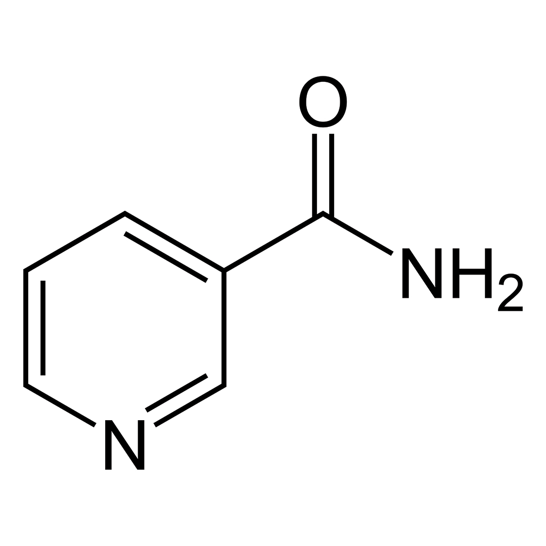
| CAS Number | 59-67-6 |
| Other Names | Nicotinic Acid, Niacin, Pyridine-3-carboxylic acid, 3-pyridinecarboxylic acid |
| IUPAC Name | pyridine-3-carboxylic acid |
| Molecular Formula | C₆H₅NO₂ |
| Molecular Weight | 123.11 |
| Purity | ≥99% Pure (LC-MS) |
| Liquid Availability | N/A |
| Powder Availability |  50 grams |
| Gel Availability | N/A |
| Storage | Store in cool dry environment, away from direct sunlight. |
| Certificate of Analysis | Due to this product’s nature, this chemical does not have a COA associated with it. |
| Terms | All products are for laboratory developmental research USE ONLY. Products are not for human consumption. |
What is Vitamin B3?
Vitamin B3, or niacin, is a vital water-soluble vitamin that supports overall health and well-being. As one of the eight B-complex vitamins, it plays an important role in various metabolic processes, especially those involved in energy production and enzyme function. Niacin comes in two primary forms: nicotinic acid and niacinamide, also known as nicotinamide; both of which serve different biological roles but fulfill the body’s need for this nutrient. Naturally present in many foods, vitamin B3 contributes to the health of the skin, nervous system, and digestive tract. A deficiency in vitamin B3 can result in pellagra, a serious condition marked by symptoms such as skin inflammation, diarrhea, and cognitive issues, underlining the nutrient’s essential role in bodily functions.
Main Research Findings
1) Application of topical vitamin B3 results in a reduced appearance of wrinkles in the skin, decreased yellowing, and improved skin elasticity.
2) Supplementation with vitamin B3 has the potential to improve inner retinal function in cases of glaucoma.
Selected Data
1) This clinical study conducted by the research team of Bissett et al investigated the effects of niacinamide, a form of vitamin B3, on facial aging and skin biology through both human trials and laboratory-based fibroblast analysis. The study was conducted with oversight to ensure compliance with the established protocol, and a statistician was responsible for reviewing the study design and analyzing the data to evaluate statistical significance. The human component of the study focused on facial aging and included 50 healthy white female participants between the ages of 35 and 60. All subjects were non-pregnant and non-lactating [1].
The design of the study was double-blind and placebo-controlled, using a split-face method with randomized left-right treatment assignment. Participants were selected based on the presence of visible signs of aging, specifically fine lines or wrinkles in the eye area and poor texture in the cheek area. Only those with a baseline score of 2.0 or greater on a 0–5 scale were eligible. Poor skin texture was defined by enlarged pores and a rough, uneven appearance often described as “pebbly” or “orange peel-like.” Additionally, subjects were required to have at least two hyperpigmented spots of at least ¼ inch in diameter on each side of the face.
Before the actual testing began, participants underwent a 2-week washout period during which they used a standardized facial cleanser and a commercial oil-in-water moisturizer twice daily. After this phase, the moisturizer was replaced with test formulations for the remainder of the 12-week study, while the cleanser remained the same. The test formulations were oil-in-water moisturizers with one version containing niacinamide and the other serving as a control, identical in every way except for the absence of niacinamide. These were distributed in blind-coded, opaque tubes labeled for use on either the left or right side of the face. Participants applied a pea-sized amount of approximately 0.4 grams, of each product to the designated side of their face twice daily: once in the morning and once in the evening, with the latter at least one hour before bedtime. New supplies of the test products were provided at baseline, and again at weeks 4 and 8 [1].
To evaluate the effects of the treatments, skin elasticity measurements were obtained using a Cutometer, which detects dermal changes with an 8 mm probe. Additionally, digital facial images were captured at baseline, and at weeks 4, 8, and 12. These images were recorded using two systems: the Wrinkle Imaging System and the Color Imaging System. The Wrinkle Imaging System used high magnification and non-polarized incandescent lighting to emphasize surface features such as fine lines and wrinkles. The Color Imaging System used low magnification and polarized fluorescent lighting to eliminate shine, allowing for precise evaluation of skin color and pigmentation. All photographs were taken under strictly controlled conditions, including standardized lighting, camera distance, and subject positioning. Prior to imaging, subjects acclimated for 30 minutes in a room with controlled temperature and humidity and were covered with black drapes to eliminate visual distractions [1].
The effectiveness of the niacinamide-containing product was assessed using both subjective and objective methods. Expert graders, trained in dermatological assessment, conducted a visual perception study. They evaluated facial images side-by-side at different time points using color-calibrated monitors. Images were randomized in terms of treatment side and presentation order. Graders selected which side appeared improved for specific skin attributes and rated the extent of improvement on a 0–4 scale. Each image pair was reviewed independently by three graders, and their scores were averaged. In addition to this visual analysis, computer-based image analysis was also employed. Proprietary algorithms were used to quantify color changes such as yellowing or hyperpigmentation, and surface characteristics such as wrinkle length and texture area.
Complementing the clinical testing, the study also included laboratory experiments using cultured human fibroblasts to evaluate biological mechanisms. Fibroblasts were obtained from neonatal tissue and a 57-year-old donor, and cultured in a minimum essential medium for varying durations. For tests involving treatment, fibroblasts from the older donor were cultured for five to seven days until they reached near-confluency. The supernatant was collected from duplicate cultures and analyzed for its content of hyaluronic acid, a major glycosaminoglycan that contributes to skin hydration and elasticity. Hyaluronic acid levels were measured using a commercial antibody-based assay kit [1].
Overall, the study was designed to comprehensively evaluate the cosmetic and biological effects of niacinamide on aging skin through both visual and quantitative assessments. The dual approach of clinical trials and culture analysis offered insight into not only the visual improvements observed with niacinamide use but also its potential mechanisms of action at the cellular level [1].
2) The study performed by the research team of Hui et al was a prospective, double-masked, randomized crossover clinical trial investigating the effects of high-dose niacinamide on retinal function in individuals with glaucoma. Eligibility criteria for the experiment included adults with early to moderate glaucoma as defined by a visual field mean deviation of ≥−10 dB and a visual acuity of at least 6/18. Reliability criteria included fewer than 33% fixation losses, false positives, and false negatives. Eligible participants needed reproducible visual field defects, defined as three neighboring points with sensitivity below age-matched norms and a probability of <2%. If both eyes met criteria, the eye with the better visual field was chosen, assuming it would be more responsive to treatment [2].
Exclusion criteria encompassed pregnancy, breastfeeding, allergy to niacinamide or niacin, a recent cancer diagnosis, apart from treated basal or squamous cell carcinoma, liver disease, or stomach ulcers. Ocular exclusions included intraocular surgery within six months, apart from uncomplicated cataract surgery within three months, and retinal conditions affecting visual function, such as age-related macular degeneration or diabetic retinopathy.
The study employed an accelerated dosing protocol to facilitate tolerability of high-dose niacinamide. Participants were randomly assigned to receive either niacinamide or a placebo in the first 12-week phase. The niacinamide treatment consisted of 1.5 g/day for six weeks followed by 3.0 g/day for another six weeks. Placebo tablets were manufactured to closely match the appearance and texture of niacinamide tablets, and all packaging was coded to maintain blinding. To preserve masking, those on placebo also doubled their tablet count at the six-week mark. Participants, physicians, assessors, and statisticians were all blinded to treatment allocations and the chosen dose was based on prior preclinical and ongoing human trials [2].
After 12 weeks, participants crossed over to the opposite treatment arm without a washout period, as it was assumed that any residual effect of niacinamide would be negligible by the next review. Participants continued their usual glaucoma medications during the study. Adherence to treatment was monitored through tablet counts at each visit, with a minimum acceptable adherence set at 70%, equivalent to no more than two missed doses per week.
Clinical assessments were conducted at baseline and every six weeks. Evaluations included visual acuity, intraocular pressure with Icare PRO rebound tonometry, blood pressure measurements, and slit lamp examination. Intraocular pressure was measured with the same device throughout the study to ensure consistency. If the device indicated measurement variability of ≥15% deviation, the test was repeated. Standard automated perimetry was used to assess visual field function. Pupil dilation was performed using 0.5% tropicamide and 2.5% phenylephrine. Electroretinography was performed under photopic conditions following at least 10 minutes of light adaptation. Custom-made DTL-like electrodes recorded electroretinography using a handheld RETeval device, with red flash stimuli on a blue background. Stimuli were calibrated using a radiometer with a photopic filter. Additionally, optical coherence tomography was used to measure circumpapillary retinal nerve fiber layer thickness [2].
The primary objective of the trial was to evaluate changes in retinal function after 12 weeks of niacinamide treatment, with a specific focus on retinal ganglion cell function. This was measured using the PhNR Vmax ratio that is the ratio of the photopic negative response (PhNR) amplitude to the b-wave amplitude. This ratio accounts for inter-session variability and has been refined in prior lab work to detect functional changes over short periods. Secondary outcomes included changes in individual electroretinography parameters, visual field indices, mean light sensitivity, retinal nerve fiber layer thickness, intraocular pressure, and mean arterial pressure [2].
Electroretinography data processing was conducted using custom MATLAB scripts by an assessor blinded to group assignments. Waveforms underwent a 0.3–300 Hz bandpass filter and third-order polynomial detrending. Amplitudes of the a-wave, b-wave, and PhNR were extracted. Amplitude responses across varying luminance levels were modeled using a saturating hyperbolic function, where Vmax represents the maximal response amplitude. The Vmax ratio was a central focus due to its sensitivity and reduced susceptibility to inter-session variability. Visual field data was analyzed in both conventional logarithmic decibel scale and converted linear units for correlation analysis. The mean light sensitivity was calculated as the mean sensitivity excluding test points adjacent to the blind spot.
Data analysis was conducted in a fully masked fashion. Placebo and niacinamide treatments were labeled as “treatment A” or “treatment B,” and the statistician received de-identified data. Treatment effects were analyzed using linear regression and adjusted for baseline values. If a significant change was observed at 12 weeks, 6-week data were also assessed. PhNR changes were also evaluated relative to previously established coefficients of repeatability including 27% for PhNR Vmax and 28% for Vmax ratio [2].
Discussion
1) The clinical evaluation of topical 5% niacinamide performed by the research team of Bissett et al revealed multiple beneficial effects on aging facial skin. These improvements were measured and observed across a range of common age-related skin concerns, including pigmentation irregularities, red blotchiness, wrinkles, yellowing, and elasticity. The treatment outcomes were consistent with findings from previous studies and supported both by subjective expert grading and objective instrumental assessments [1]
One of the most noticeable benefits of niacinamide treatment was a reduction in hyperpigmented spots, commonly associated with age and sun exposure. These spots, often appearing as lentigines or dark patches, were significantly diminished after eight weeks of twice-daily application of the niacinamide-containing moisturizer. The effect was consistent with prior studies on different skin types, including white and Japanese facial skin. This suggests that niacinamide’s depigmenting effect is reliable across ethnicities, highlighting its broad applicability in cosmetic skincare.
In addition to improving pigmentation irregularities, niacinamide was effective in reducing red facial blotchiness, another common sign of aging or irritation. Red blotchiness was gradually reduced over the course of the 12-week study, with significant differences from baseline observed at the final time point. This improvement in skin color uniformity is consistent with earlier clinical studies and adds to the evidence supporting niacinamide’s anti-inflammatory or barrier-repairing properties [1].
A particularly important result from this study was the reduction in fine lines and wrinkles, which was observed after eight weeks of treatment. Wrinkle depth and visibility were evaluated using high-resolution imaging and expert grading, and a statistically significant decrease in these features was documented. Two biological mechanisms may explain this anti-wrinkle effect. First, niacinamide appears to stimulate collagen production. Collagen is a critical structural protein that maintains skin firmness and elasticity, and its loss contributes to wrinkle formation. Prior work involving cultured fibroblasts confirmed this collagen-boosting property [1].
Second, niacinamide may help reduce the accumulation of excess dermal glycosaminoglycans. Although glycosaminoglycans such as hyaluronic acid play essential roles in hydration and tissue structure, excessive levels can disrupt dermal architecture in aging skin. Cell culture experiments using fibroblasts from both neonatal and 57-year-old donors showed that older fibroblasts produced two to three times more glycosaminoglycans than younger ones. This age-related increase in glycosaminoglycans production was mitigated by treatments with niacinamide, as well as by all-trans retinoic acid, a well-established anti-aging compound used as a positive control. Thus, the wrinkle reduction observed clinically may be due to a combination of enhanced collagen synthesis and better regulation of glycosaminoglycan levels in the dermis [1].
Another hallmark of aging skin is yellowing, also referred to as sallowness, which gives the skin a dull and unhealthy appearance. This study evaluated skin yellowing using objective color measurements that quantifies yellow tones. Niacinamide was effective in preventing an increase in skin yellowing over the 12-week study period. Although the exact biological mechanism behind this effect is not fully understood, one proposed explanation involves the prevention of protein glycation. Glycation is a non-enzymatic chemical reaction that leads to the formation of advanced glycation end-products, which can discolor proteins like collagen and elastin, leading to a yellowish appearance. Niacinamide may interfere with this process, though further investigation is needed to confirm this mechanism.
Improvements in skin elasticity were also documented through cutometry, an instrumental method that measures mechanical properties of the skin. Two key cutometry parameters were evaluated: R5, which represents the viscoelastic properties of the skin, and R7, which measures the skin’s ability to return to its original state after deformation. Both parameters showed significant improvements after 12 weeks of niacinamide treatment, indicating that the skin had become more supple and resilient. Although the precise biochemical pathways responsible for these changes are not entirely clear, they may be linked to the same mechanisms involved in wrinkle reduction including: dermal remodeling through increased collagen production and reduced glycation of dermal matrix components such as collagen and elastin [1].
In conclusion, the clinical use of topical 5% niacinamide over a 12-week period demonstrated a broad spectrum of benefits for aging skin. These included significant reductions in hyperpigmented spots, red blotchiness, and wrinkles; prevention of yellowing; and measurable improvements in skin elasticity. The mechanisms underlying these effects likely involve a combination of anti-inflammatory activity, enhanced extracellular matrix remodeling, stimulation of collagen synthesis, regulation of glycosaminoglycan production, and potential anti-glycation effects. These findings strongly support the use of niacinamide as an effective, multi-functional ingredient in topical treatments designed to address the visible signs of facial skin aging [1].
2) The study conducted by Hui et al examined the effects of high-dose nicotinamide on retinal function and visual field measures in patients with glaucoma. Among 94 individuals screened, 57 were enrolled and randomized into the trial and 86% completed the study. The average age of participants was approximately 65.5 years, and 39% were female. The treatment and placebo groups were balanced with no significant differences in their glaucoma therapy prior to or during the study. 70% of participants had primary open-angle glaucoma, while 16% had chronic angle-closure glaucoma. Adherence to the treatment protocol was high, with over 94% of participants complying with both the low- and high-dose nicotinamide regimens, indicating the treatment’s good tolerability. Only two participants were deemed non-adherent during the nicotinamide phase due to missed visits and were excluded from the final analysis [2].
The primary objective was to evaluate changes in retinal ganglion cell function, specifically using electroretinographic markers such as the photopic negative response Vmax and Vmax ratio. After 12 weeks of high-dose nicotinamide, significant improvements in these inner retinal function markers were observed. Specifically, 23% of participants on nicotinamide showed improvement in PhNR Vmax that exceeded the 95% coefficient of repeatability, while only 9% on placebo reached this threshold. For the Vmax ratio, 21% on nicotinamide improved beyond the coefficient of repeatability, compared to 14% on placebo. A small number of participants experienced deterioration in these markers, with 7–9% on nicotinamide and 5–9% on placebo showing reductions.
The overall treatment effect between nicotinamide and placebo was statistically significant for both PhNR Vmax and the Vmax ratio. Relative to baseline, there was a 12.6% improvement in Vmax ratio with nicotinamide, compared to a 3.6% improvement with placebo. For PhNR Vmax, nicotinamide led to a 14.8% improvement versus 5.2% with placebo. Importantly, no carryover or period effects were detected, validating the crossover design. Sensitivity analyses excluding participants with missing electroretinographic data confirmed the robustness of these findings. Improvements were only observed after the full 12-week high-dose nicotinamide regimen, with no significant changes detected at the 6-week, lower-dose stage [2].
No significant treatment effects were observed for outer retinal function as measured by electroretinographic a-waves as a measure of photoreceptor function and b-wave as a measure of bipolar cell function amplitudes or their implicit times. Furthermore, no differences were seen in the parameters used to fit the b-wave response between the nicotinamide and placebo groups, indicating that the effects of nicotinamide were likely specific to inner retinal ganglion cell function [2].
In terms of visual field outcomes, global indices such as mean deviation and pattern standard deviation did not differ significantly between treatment arms. After 12 weeks, mean deviation was 0.10 dB higher in the nicotinamide group compared to placebo, and pattern standard deviation was slightly lower by 0.25 dB, both of which were not statistically significant. However, when evaluating individual-level changes, a higher proportion of participants on nicotinamide experienced a clinically relevant improvement in mean deviation compared to placebo of 27% vs 16%, respectively. Conversely, fewer nicotinamide-treated participants showed a ≥1 dB decline of 4% vs 12%. This pattern was not observed for pattern standard deviations, where changes were similar across both groups.
Further supporting the relevance of the electroretinography findings, changes in PhNR Vmax correlated moderately with changes in visual field mean deviation at 12 weeks in both treatment arms. A similar moderate positive correlation was also found between PhNR Vmax and mean light sensitivity in both groups. These associations suggest that improvements in retinal ganglion cell function may relate to improved visual field performance [2].
Secondary outcome measures such as intraocular pressure, mean arterial pressure, and visual acuity showed no significant differences between nicotinamide and placebo. The mean intraocular pressure following nicotinamide treatment was 13.8 mmHg compared to 13.4 mmHg on placebo, with a non-significant mean difference of 0.2 mmHg. There were also no changes in average retinal nerve fiber layer thickness after NAM treatment, indicating that structural changes did not accompany the functional improvements within the short study period [2].
In conclusion, this trial demonstrates that high-dose nicotinamide can improve retinal ganglion cell function in patients with glaucoma without causing significant adverse effects. While global visual field measures did not change significantly, more participants on nicotinamide experienced meaningful improvements in visual field mean deviation, and electroretinography improvements correlated positively with visual field changes. These findings suggest that nicotinamide may offer a safe and effective adjunctive strategy to support inner retinal function in glaucoma, though longer-term studies are needed to determine structural benefits and sustained visual outcomes [2].
Disclaimer
**LAB USE ONLY**
*This information is for educational purposes only and does not constitute medical advice. THE PRODUCTS DESCRIBED HEREIN ARE FOR RESEARCH USE ONLY. All clinical research must be conducted with oversight from the appropriate Institutional Review Board (IRB). All preclinical research must be conducted with oversight from the appropriate Institutional Animal Care and Use Committee (IACUC) following the guidelines of the Animal Welfare Act (AWA).
Citations
[1] Bissett DL, Oblong JE, Berge CA. Niacinamide: A B vitamin that improves aging facial skin appearance. Dermatol Surg. 2005;31(7 Pt 2):860-865. doi:10.1111/j.1524-4725.2005.31732
[2] Hui F, Tang J, Williams PA, et al. Improvement in inner retinal function in glaucoma with nicotinamide (vitamin B3) supplementation: A crossover randomized clinical trial. Clin Exp Ophthalmol. 2020;48(7):903-914. doi:10.1111/ceo.13818
Vitamin B3 is sold for laboratory research use only. Terms of sale apply. Not for human consumption, nor medical, veterinary, or household uses. Please familiarize yourself with our Terms & Conditions before ordering.



Reviews
There are no reviews yet.