Main Research Findings
1) Use of VIP in post-refractive eye surgery was shown to indicate correlation between postoperative dry eye and optic nerve damage, as well as relieve postoperative dry eye symptoms.
2) Treatment with VIP was found to improve the likelihood of survival from respiratory failure after 60 days in cases of critical COVID 19.
Selected Data
1) The study completed by the research team of Kang et al was designed as a prospective, non-randomized, longitudinal cohort study to investigate the changes in tear Vasoactive Intestinal Peptide (VIP) concentration following two common types of laser keratorefractive surgery: Laser-Assisted Sub-Epithelial Keratomileusis (LASEK) and Femtosecond Laser-Assisted In Situ Keratomileusis (FS-LASIK). The primary objectives were to explore the temporal dynamics of tear VIP, its association with postoperative dry eye symptoms, and the factors influencing these outcomes in different surgical groups [1].
A total of 45 subjects were recruited, comprising 23 individuals undergoing LASEK and 22 undergoing FS-LASIK. The choice of surgical procedure was based on individual patient preference and surgical indications, thus explaining the non-randomized nature of the study. Inclusion criteria to ensure standardization were as follows: participants were between 18 and 45 years old, had stable refractive error for at least two years (defined as <0.25 D change per year), possessed an estimated residual stromal bed thickness of not less than 280 µm, and for those with spherical equivalent exceeding -5.00D, only FS-LASIK was an option. Exclusion criteria precluded individuals with topography consistent with keratoconus, ocular comorbidities, prior ophthalmic surgery, systemic disorders, relevant medication history (especially growth inhibitors and glucocorticoids), pregnancy, or monovision, to minimize confounding factors.
Both LASEK and FS-LASIK surgeries were performed by the same experienced surgeon to maintain consistency. For LASEK, a corneal epithelial flap was created using a 20% alcohol solution instilled within a trephine centered on the pupil, leaving a superior hinge. The ablation was then performed using an EX500 excimer laser with an optical diameter of 6.25 to 6.75 mm and a 1.5-mm transition zone. Mitomycin C was explicitly not used in any LASEK cases. A bandage contact lens was inserted post-LASEK for 7 days. In FS-LASIK, a corneal flap was created using an Allegretto Wave-Light FS-200 laser with specific parameters: 110 µm depth, 8.5–9.1 mm diameter, a 105° superior gas canal in the right eye, and a 75° superior gas canal in the left eye. The ablation rate for FS-LASIK was 500 kHz, with a pulse energy of approximately 160n [1].
Detailed systemic and ocular disease histories were meticulously collected from all subjects. Postoperative management included a tapered regimen of topical steroid (Fluorometholone 0.1%) for 4 months in LASEK and over 12 days in FS-LASIK, topical antibiotic (Tobramycin 0.003%) for 7 days, and topical lubricant eye drops (HYCOSAN) for approximately 3 months. Follow-up examinations, conducted by the same surgical team to ensure standardization, included measurements of uncorrected distance visual acuity, slit-lamp examination, intraocular pressure (IOP), and corneal topography.
Outcome measures were employed to comprehensively assess ocular health and VIP levels at multiple time points: before surgery, and at 1 day, 1 week, and 1 month post-surgery. Tear samples for VIP concentration analysis were collected by inserting dry sharp-tip microsponges into the inferior conjunctival fornix for 30 seconds, followed by storage at -80 °C. These samples were subsequently diluted with phosphate-buffered saline, and protein concentration was determined using a BCA Protein Assay Kit. Tear VIP concentration was quantified using a specific enzyme-linked immunosorbent assay (ELISA) kit, with a detection limit of 4.69 ng/ml. Dry eye indices included tear-film breakup time (TBUT), measured as the time from a complete blink to the first break in the tear film (averaged over three repetitions), ocular staining (assessed using the Oxford grading scale), and the Ocular Surface Disease Index (OSDI) questionnaire (Chinese version). Corneal parameters such as the thinnest corneal thickness (CCT) were measured thrice using Pentacam (Oculus GmbH), and corneal ablation depth (AD) and predicted residual corneal thickness (RCT) were determined by the Allegretto EX500 excimer laser software [1].
All statistical analyses were performed using SPSS software. Continuous variables were expressed as mean ± SD or median (interquartile range [IQR]) and tested for normal distribution. Independent sample t-tests compared basic surgical parameters between groups. Nonparametric tests were used for changes in tear VIP and dry eye indices over time, and Mann–Whitney U tests for between-group comparisons of these indices. Pearson and Spearman correlation tests were used to examine relationships between VIP concentration and dry eye symptoms, with coefficients ranking dry eye factor correlation [1].
2) The study performed by researchers Youssef et al was designed as a multicenter, randomized, double-blind, placebo-controlled phase 2b/3 clinical trial. Its primary objective was to evaluate the efficacy and safety of intravenous Aviptadil (synthetic VIP) in improving survival and recovery from respiratory failure in critically ill COVID-19 patients. Participants were eligible if they were 18 years or older, had confirmed critical COVID-19 by polymerase chain reaction, and required significant respiratory support, specifically high-flow nasal cannula (HFNC) greater than 20L, noninvasive ventilation (NIV), or mechanical ventilation (MV). Exclusion criteria included factors such as active bacterial/fungal infections, recent organ transplantation, certain autoimmune diseases, recent use of specific immunosuppressants, or an active DNR/DNI order, ensuring a focused and manageable patient cohort [2]
Participants were randomized in a 2:1 ratio to receive either Aviptadil or placebo. Aviptadil was administered as three 12-hour intravenous infusions over three days, with graduating doses of 50, 100, and 150 pmol/kg/hr. The placebo group received normal saline administered identically. A robust blinding strategy was implemented, with only an unblinded research pharmacist preparing the identically labeled infusions, ensuring that study personnel, patients, and families remained unaware of treatment assignment.
The primary endpoint of the study, as modified during the trial, was “alive at day 60 and free of respiratory failure,” defined as being alive, discharged from acute care, and no longer requiring more than 2L of low-flow oxygen. The key secondary endpoint was survival at 60 days post-treatment. To assess biological response, two intermediate endpoints were prespecified: changes in Respiratory Distress Ratio (RDR), defined as Pao2:Fio2 (P:F) ratio, and serum levels of inflammatory cytokines, specifically interleukin-6 (IL-6), measured daily through day 7. Disease severity at baseline was assessed using the National Institute of Allergy and Infectious Diseases (NIAID) 8-point ordinal scale. Statistical analyses were performed on a modified intention-to-treat (mITT) population [2].
Logistic regression was used for binary endpoints (primary and secondary), controlling for baseline ventilation status. Mixed-model repeated measure (MMRM) was applied to analyze daily changes in NIAID score, RDR, and cytokine levels. Preplanned subgroup analyses considered baseline ventilation status and concurrent medications, while a post-hoc analysis examined the effect of the site of care (tertiary vs. community hospital) due to observed mortality differences. The study’s power calculation underwent adjustments, increasing the sample size to 198 subjects to achieve 81% power to detect a 22% absolute increase in respiratory failure resolution. All statistical procedures and protocol modifications, including endpoint reversal from the original design, were done in consultation with the FDA and DMC, ensuring rigor and responsiveness to emerging data in the rapidly evolving COVID-19 landscape [2].
Discussion
1) The findings of the study performed by Kang et al revealed significant changes in tear Vasoactive Intestinal Peptide (VIP) concentrations and dry eye indices following LASEK and FS-LASIK surgeries, along with crucial correlations that shed light on the neuroimmune response to refractive surgery. A total of 23 subjects underwent LASEK, and 22 subjects underwent FS-LASIK. Baseline surgical parameters showed notable differences between the two groups due to the study’s inclusion criteria; specifically, spherical equivalent (SE), corneal ablation depth (AD), and predicted residual corneal thickness (RCT) differed significantly. This was expected, as patients with higher myopia were directed towards FS-LASIK [1].
A key finding was the significant increase in tear VIP concentration post-surgery in both LASEK and FS-LASIK groups, reaching its peak approximately one week after the procedure. This elevation in VIP suggests a robust neuroimmune response triggered by the surgical trauma. Notably, there was no significant difference in tear VIP concentrations between the LASEK and FS-LASIK groups at any measured time point, indicating a general response regardless of the specific surgical technique.
Dry eye symptoms, as assessed by TBUT, corneal staining, and OSDI, also showed significant changes. In the LASEK group, TBUT decreased, and corneal staining and OSDI increased significantly one week post-surgery, with the increase in OSDI and corneal staining also observed at 1 day post-surgery. For the FS-LASIK group, TBUT decreased significantly at 1 month, while OSDI increased significantly at both 1 week and 1 month post-surgery. Comparing the two surgical groups, LASEK subjects generally experienced more severe dry eye indices and a greater degree of deterioration after 1 week compared to FS-LASIK subjects, with corneal staining showing a significant difference. At 1 month post-surgery, OSDI scores were significantly higher in the FS-LASIK group than in the LASEK group [1].
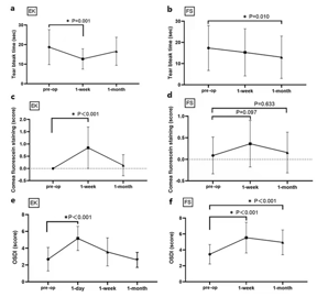
Figure 1: Changes in (A, B) TBUT, (C, D) cornea fluorescein staining, and (E, F) OSDI score, measured pre-op, and at 1 and 3 weeks.
Crucial correlation analyses revealed that increases in VIP concentrations post-surgery were negatively correlated with dry eye indices. Specifically, in the LASEK group, VIP3-1 (change in VIP from pre-op to 1 week) was negatively correlated with TBUT1-3 (change in TBUT), corneal staining3 (change in corneal staining), and OSDI3 (change in OSDI). A similar negative correlation was observed between VIP4-1 (change in VIP from pre-op to 1 month) and TBUT1-4. In the FS-LASIK group, VIP3-1 also showed negative correlations with TBUT1-3, corneal staining3, and OSDI3, while VIP4-1 was negatively correlated with TBUT1-4 and corneal staining4. These findings indicate that higher increases in VIP concentration are associated with better dry eye outcomes [1].
A significant negative correlation was found between VIP3-1 and corneal AD in both LASEK and FS-LASIK groups. This implies that a greater corneal ablation depth is associated with a lower increase in VIP concentration, suggesting that the extent of nerve damage (reflected by AD) can modulate the VIP response. No other factors such as age, IOP, CCT, RCT, or haze were found to correlate with tear VIP concentration. Furthermore, no correlation was identified between preoperative tear VIP concentration and dry eye symptoms, indicating that the VIP response is primarily a reaction to the surgical event rather than a baseline state.
In summary, the study concludes that refractive surgery acts as a trauma model, triggering an emergency protective response that mobilizes tear VIP. This elevated VIP concentration, especially within the first week, correlates with reduced dry eye symptoms. The negative correlation between VIP increase and corneal ablation depth highlights the link between surgical trauma, nerve injury, and the VIP response. These findings emphasize VIP’s neuroimmune role in regulating adverse injury stimulation and provide valuable insights into the pathogenesis and potential treatment strategies for postoperative dry eyes [1].
2) The clinical trial conducted by Youssef et al investigated Aviptadil (synthetic VIP) for critical COVID-19 respiratory failure, ultimately enrolled 203 eligible participants between May and December 2020, with 131 receiving Aviptadil and 65 receiving placebo, analyzed under a modified intention-to-treat population. Baseline characteristics indicated a more severe disease state in the Aviptadil group, particularly regarding the proportion of patients on mechanical ventilation (27.5% vs 16.9%), underscoring the importance of controlling for baseline ventilation status in the analyses. Treatment adherence was high, with 91% of Aviptadil recipients completing all three infusions; dose adjustments were primarily due to mild-to-moderate diarrhea, a known side effect of VIP [2].
The primary endpoint, “alive at day 60 and free from respiratory failure,” did not reach statistical significance when controlling for baseline ventilation status. This outcome was largely influenced by 10 Aviptadil-treated patients who, though alive at day 60, remained on ongoing ventilator support in rehabilitation centers, not meeting the “free from respiratory failure” criterion despite clinical improvement. However, the key secondary endpoint, survival at 60 days, demonstrated a statistically significant improvement with Aviptadil, across all patients and sites of care. This survival benefit was particularly pronounced in the most severely ill subgroup, defined by a NIAID score 2, indicating need for mechanical ventilation at baseline, where Aviptadil treatment was associated with a 10-fold increased odds of survival compared to placebo. Additionally, a statistically significant hazard ratio for survival through 60 days was observed, even when controlling for baseline ventilation [2].
Regarding biological endpoints, Aviptadil exhibited significant effects on respiratory distress and inflammation. The Respiratory Distress Ratio (RDR) showed significant improvement with Aviptadil treatment across the first seven days following administration, with particularly notable differences in the HFNC subgroup. In terms of inflammatory markers, Aviptadil significantly prevented the rise of serum interleukin-6 (IL-6) levels from day 3 to day 7 compared to placebo, with independent significance observed on day 3 and day 7. Higher IL-6 levels were found to be highly predictive of a failure to achieve the primary endpoint and overall survival, suggesting that Aviptadil’s anti-inflammatory action contributed to patient outcomes. A subgroup analysis further revealed a significant advantage for Aviptadil in patients who developed respiratory failure despite prior remdesivir treatment, showing a 3.1-fold increased odds of achieving the primary endpoint and a 4.15-fold increased odds of survival.
The safety profile of Aviptadil was favorable. A post-hoc analysis incorporating the site of care as a covariate revealed a significant interaction between treatment and hospital type, with higher mortality rates observed in community hospitals, a finding attributed to the COVID-19 surge and resource limitations during the study period. Despite the failure to meet the prespecified definition of the primary endpoint due to specific classifications of recovery, Aviptadil consistently demonstrated improved survival, enhanced lung oxygenation, and attenuated cytokine storm across varying disease severities and hospital settings, supporting its beneficial biological effects in critical COVID-19 [2] .
Disclaimer
**LAB USE ONLY**
*This information is for educational purposes only and does not constitute medical advice. THE PRODUCTS DESCRIBED HEREIN ARE FOR RESEARCH USE ONLY. All clinical research must be conducted with oversight from the appropriate Institutional Review Board (IRB). All preclinical research must be conducted with oversight from the appropriate Institutional Animal Care and Use Committee (IACUC) following the guidelines of the Animal Welfare Act (AWA).
Citations
[1] Kang Y, Hu Q, Li X, Guo Z, Wu Q, Zhang H. Role of tear vasoactive intestinal peptide on dry eyes after laser keratorefractive surgery. BMC Ophthalmol. 2023;23(1):167. Published 2023 Apr 20. doi:10.1186/s12886-023-02857-w
[2] Youssef JG, Lavin P, Schoenfeld DA, et al. The Use of IV Vasoactive Intestinal Peptide (Aviptadil) in Patients With Critical COVID-19 Respiratory Failure: Results of a 60-Day Randomized Controlled Trial. Crit Care Med. 2022;50(11):1545-1554. doi:10.1097/CCM.0000000000005660









 5 milligrams vial, 10 milligrams vial (lyophilized/freeze-dried)






Reviews
There are no reviews yet.